You have no items in your shopping cart.
Cart summary
Item 1 of 6
Item 1 of 6
Myosin phospho S19 antibody
Catalog Number: orb345407

| Catalog Number | orb345407 |
|---|---|
| Category | Antibodies |
| Description | Myosin phospho S19 antibody |
| Species/Host | Rabbit |
| Clonality | Polyclonal |
| Tested applications | ELISA, IHC, IP, WB |
| Reactivity | Human, Mouse |
| Isotype | IgG |
| Immunogen | Human Myosin Light Chain phospho peptide corresponding to a region near the amino terminus of the human smooth/non-muscle form of myosin regulatory light chain conjugated to Keyhole Limpet Hemocyanin (KLH). |
| Concentration | 1.0 mg/mL |
| Dilution range | ELISA: 1:10,000 - 1:30,000, IHC: 2.5 µg/ml, IP: 1:100, WB: 1:1,000 - 1:5,000 |
| Form/Appearance | Liquid (sterile filtered) |
| Purity | This affinity purified antibody is directed against the regulatory light chain of smooth and non-muscle myosin. The antibody is phosphospecific and detects monophosphorylated and diphosphorylated forms of the protein. The product was affinity purified from monospecific antiserum by immunoaffinity purification. Antiserum was first purified against the phosphorylated form of the immunizing peptide. The resultant affinity purified antibody was then cross-adsorbed against the non-phosphorylated form of the immunizing peptide. This phosphospecific polyclonal antibody is specific for the phosphorylated pS19/pS20 form of the protein, depending on the source origin of the protein. Reactivity with non-phosphorylated myosin light chain is less than 1% by ELISA. Cross reactivity is expected with myosin light chain from human and mouse. Reactivity with the protein from other species has not been determined. However, the sequence of the immunogen is nearly identical in mammalian and avian species. BLAST search analysis was used to determine that the smooth and non-muscle forms of myosin regulatory light chain have identical sequences. Cross reactivity is expected. |
| Conjugation | Unconjugated |
| UniProt ID | P19105 |
| NCBI | AAH16372.1 |
| Storage | Store vial at -20° C or below prior to opening. This vial contains a relatively low volume of reagent (25 µL). To minimize loss of volume dilute 1:10 by adding 225 µL of the buffer stated above directly to the vial. Recap, mix thoroughly and briefly centrifuge to collect the volume at the bottom of the vial. Use this intermediate dilution when calculating final dilutions as recommended below. Store the vial at -20°C or below after dilution. Avoid cycles of freezing and thawing. |
| Buffer/Preservatives | 0.01% (w/v) Sodium Azide |
| Alternative names | rabbit anti-Myosin p19/pS20 antibody, Myosin regul Read more... |
| Note | For research use only |
| Application notes | This phospho specific polyclonal antibody was tested by ELISA, immunohistochemistry, and immunoblotting. Immunoblotting was used to show reactivity with unstimulated and stimulated cardiac myocytes. The antibody was also reactive with the phosphorylated form of the immunizing peptide and minimally reactive with the non-phosphorylated form of the immunizing peptide. Although not tested, this antibody is likely functional by immunoprecipitation. |
| Expiration Date | 12 months from date of receipt. |
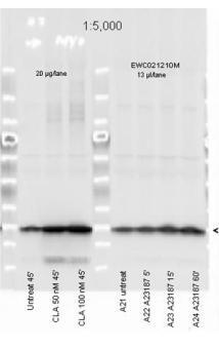
Affinity Purified Phospho specific antibody to Monophosphorylated Regulatory Light Chain of Smooth and Non-muscle Myosin at pS19/pS20 was used at a 1:5000 dilution to detect myosin light chain by Western blot. Either 13 µL or 20 µg of a mouse cardiac myocyte lysate was loaded on a 4-20% Criterion gel for SDS-PAGE. Samples were either mock-treated or CLA-treated, as indicated. After washing, a 1:5000 dilution of HRP conjugated Gt-a-Rabbit IgG (orb347654) preceded color development using Amersham's substrate system. Other detection methods will yield similar results.

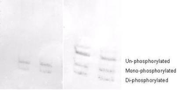
Affinity purified phosphospecific antibody to phosphorylated regulatory light chain of smooth and non-muscle Myosin at pS19/pS20 was used at a 1:1000 dilution to detect myosin light chain by Western blot on 3T3 cell lysates. A standard urea/glycerol gel without SDS was used to separate phospho forms of regulatory light chain according to mass to charge ratios. In Panel A on the left, reactivity of Biorbyt's phosphospecific antibody is shown. In Panel B on the right, reactivity of commercially available pan reactive antibody that detects both un-phosphorylated and phosphorylated forms of regulatory light chain is shown. Biorbyt's phosphospecific antibody detects both mono-phosphorylated (pSer20 Mono-P-RLC) and di-phosphorylated (pThr19-pSer20 Di-P-RLC) regulatory light chain.

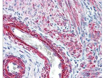
Biorbyt's affinity purified anti-Monophosphorylated RLC Smooth and Non-Muscle Myosin pS19/20 antibody was used at 2.5 µg/ml to detect signal in a variety of tissues including multi-human, multi-brain and multi-cancer slides. This image shows strong staining of both vascular and myometrial smooth muscle cells of the uterus. Tissue was formalin-fixed and paraffin embedded. The image shows localization of the antibody as the precipitated red signal, with a hematoxylin purple nuclear counterstain.
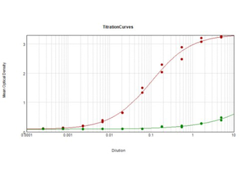
ELISA Results of Rabbit Anti-Myosin pS19/pS20 Antibody tested against BSA-conjugated peptide of immunizing peptide. Each well was coated in duplicate with 0.1 µg of Myosin pS19/pS20 [Red Line] and Myosin S19/S20 [Green Line]. The starting dilution of antibody was 5 µg/ml and the X-axis represents the Log10 of a 3-fold dilution. This titration is a 4-parameter curve fit where the IC50 is defined as the titer of the antibody. Assay performed using Goat anti-Rabbit IgG Antibody Peroxidase Conjugated (Min X Bv Ch Gt GP Ham Hs Hu Ms Rt & Sh Serum Proteins) (p/n orb347654) and TMB ELISA Peroxidase Substrate (p/n orb348651).

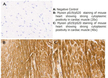
Immunohistochemistry with anti-myosin pS19/pS20 antibody showing strong cytoplasmic staining of myocytes in mouse heart muscle 20x and 40x (B & C). Formalin fixed/paraffin embedded tissue sections were subjected to antigen retrieval and then incubated with rabbit anti-myosin pS19/pS20 antibody at 1:100 dilution for 60 minutes. Biotinylated Anti-rabbit secondary antibody was used to detect primary antibody. The reaction was developed using streptavidin-HRP conjugated compact polymer system and visualized with chromogen substrate, 3'3-diamino-benzidine substrate (DAB). The sections were then counterstained with hematoxylin to detect cell nuclei.

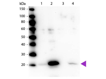
Western blot of Rabbit Anti-Myosin pS19/pS20 primary antibody. Lane 1: Regulatory Light Chain Non-Phospho recombinant protein. Lane 2: Regulatory Light Chain Phospho recombinant protein. Lane 3: Smooth Muscle Non-Phospho recombinant protein. Lane 4: Smooth Muscle Phospho recombinant protein. Load: 50 ng per lane. Primary antibody: Myosin pS19/pS20 primary antibody at 1:1000 overnight at 4°C. Secondary antibody: Peroxidase rabbit secondary antibody at 1:40000 for 60 min at RT. Blocking: orb348637 for 30 min at RT. Predicted/Observed size: 20 kDa, 20 kDa for Regulatory Light Chain Phospho. Other band(s): None.
- Item 1 of 6
Myosin phospho S19 antibody [orb345406]
ELISA, IHC, IP, WB
Human, Mouse
Rabbit
Polyclonal
Unconjugated
100 μg 
MRCL3 (phospho-Thr17+Ser18) antibody (HRP) [orb503802]
ICC, IHC-Fr, IHC-P
Bovine, Canine, Equine, Gallus, Human, Mouse, Porcine, Sheep, Zebrafish
Rat
Rabbit
Polyclonal
HRP
100 μlMRCL3 (phospho-Thr17+Ser18) antibody [orb185225]
ICC, IF, IHC-Fr, IHC-P
Bovine, Canine, Equine, Gallus, Human, Mouse, Porcine, Sheep, Zebrafish
Rat
Rabbit
Polyclonal
Unconjugated
200 μl, 100 μl, 50 μlMYL9 (phospho-S19) antibody [orb644743]
WB
Human, Mouse, Rat
Rabbit
Polyclonal
Unconjugated
25 μg, 50 μg, 100 μg, 200 μg


Submit a review
Filter by Rating

- 5 stars

- 4 stars
- 3 stars
- 2 stars
- 1 stars
