You have no items in your shopping cart.
Cart summary
Item 1 of 5
Item 1 of 5
MYC Epitope Tag antibody
Catalog Number: orb345393

| Catalog Number | orb345393 |
|---|---|
| Category | Antibodies |
| Description | MYC Epitope Tag antibody |
| Species/Host | Rabbit |
| Clonality | Polyclonal |
| Tested applications | ELISA, IHC, WB |
| Isotype | IgG |
| Immunogen | This antibody was purified from whole rabbit serum prepared by repeated immunizations with Myc epitope tag peptide, E-Q-K-L-I-S-E-E-D-L, conjugated to KLH using maleimide. The sequence corresponds to amino acids 410-419 of human c-Myc. |
| Concentration | 1.0 mg/ml |
| Dilution range | ELISA: 1:135,000, IHC: User Optimized, WB: 1:500 - 1:5,000 |
| Form/Appearance | Liquid (sterile filtered) |
| Purity | This affinity purified antibody is directed against human c-Myc and is useful in determining its presence in various assays. This polyclonal anti-Myc-tag antibody detects overexpressed proteins containing the Myc epitope tag. The antibody recognizes the Myc-tag (Glu-Gln-Lys-Leu-Ile-Ser-Glu-Glu-Asp-Leu) fused to either the amino- or carboxy- termini of targeted proteins in transfected or transformed cells. |
| Conjugation | Unconjugated |
| Storage | Store vial at -20° C prior to opening. Aliquot contents and freeze at -20° C or below for extended storage. Avoid cycles of freezing and thawing. Centrifuge product if not completely clear after standing at room temperature. This product is stable for several weeks at 4° C as an undiluted liquid. Dilute only prior to immediate use. |
| Buffer/Preservatives | 0.01% (w/v) Sodium Azide |
| Alternative names | Rabbit anti-MYC Epitope Tag Antibody, Rabbit anti- Read more... |
| Note | For research use only |
| Application notes | Anti-Myc has utility to detect the fusion protein of the Myc epitope cloned along with the target gene. As such, anti-Myc/Myc can be used to identify fusion proteins containing the Myc epitope. The antibody recognizes the Myc tag fused either to the AMINO- or CARBOXY- termini of targeted proteins. This antibody was tested by ELISA and western blotting and was tested against both the immunizing peptide and Myc-tagged recombinant proteins. Although not tested, this antibody is likely functional for immunoprecipitation and immunocytochemistry. |
| Expiration Date | 12 months from date of receipt. |
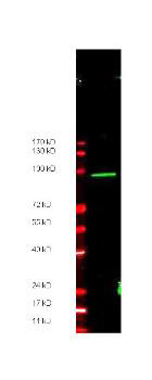
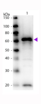
Anti-Myc epitope tag polyclonal antibody detects ~ 100 kDa CARBOXY terminal linked Myc-tagged recombinant protein present in ~35 µg of lysate by western blot.
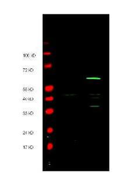
Anti-Myc epitope tag polyclonal antibody detects both AMINO and CARBOXY terminal linked Myc-tagged recombinant proteins by western blot. Polyclonal rabbit host anti-Myc epitope tag antibody was diluted to 1.0 µg/ml to detect either recombinant protein. 4-20% gradient gels were used to resolve the proteins by SDS-PAGE. The proteins were transferred to nitrocellulose using standard methods. After blocking, the membranes were probed with the primary antibody overnight at 4°C followed by washes and reaction with a 1:10000 dilution of IRDye® 800 conjugated Gt-a-Rabbit IgG (H&L) MX10 for 45 min at room temperature (Green, 800 nm channel). Pre-stained molecular weight markers are also shown (lane M, Red, 700 nm channel).
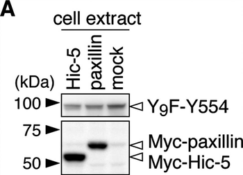
Git1 phosphorylation at Tyr-554 was enhanced by co-expression of paxillin.A, Western blotting of protein expression levels in HEK293T cells exogenously expressing the FLAG-tagged Git1-Y9F-Y554 mutant together with Myc-tagged paxillin, Myc-tagged Hic-5, or a control mock. B, Tyrosine phosphorylation of Y9F-Git1 proteins in anti-FLAG immunoprecipitates. The lower graph shows the densitometric analysis of the Western blotting data. Data are the mean ± S.E. (error bars; n=3). *, P < 0.05 significantly different from Hic-5-transfected cells by ANOVA with Fisher's PLSD post hoc tests.
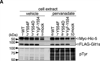
Git1 phosphorylation at Tyr-554 weakened its association with Hic-5.A, Western blotting of protein expression levels, and tyrosine phosphorylation of all proteins in HEK293T cells expressing FLAG-tagged Git1 proteins together with Myc-tagged Hic-5. Cells were treated with 100 μM pervanadate or vehicle for 15 min, and then analyzed by Western blotting using anti-FLAG M2, anti-Myc 9E10, or anti-phosphotyrosine PY20. B, Co-immunoprecipitaion of Git1 mutants with Hic-5. The immunoprecipitates from cell extracts with anti-FLAG beads were analyzed by Western blotting with an anti-FLAG or anti-Myc antibody. To verify the tyrosine phosphorylation of FLAG-tagged Git1 proteins, the same membrane was reacted with anti-phosphotyrosine PY20. Ig, immunoglobulin. The lower part shows the densitometric analysis of the relative amount of Myc-Hic-5 to FLAG-Git1 in the immunoprecipitates. Data are the mean ± S.E. (error bars; n=3). **, P < 0.01 significantly different from the wild-type with the same treatment; #, P < 0.05 or ##, P < 0.01 significant difference between vehicle- and pervanadate-treated groups by ANOVA with Fisher's PLSD post hoc tests.
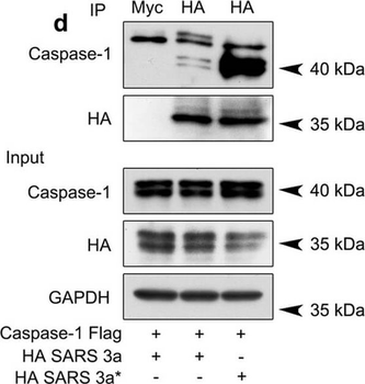
SARS 3a induces NLRP3 inflammasome activation by multiple mechanisms. A) Immunoblot analysis of the pro- and cleaved forms of caspase-1 and IL-1β after reconstitution of inflammasome in HEK 293T cells transfected with SARS 3a with or without NEK7 shRNA. B) Immunoblot analysis of the pro- and cleaved forms of caspase-1 and IL-1β after reconstitution of inflammasome and transfection with SARS 3a or SARS 3a C133A. C) Immunoblot analysis of the pro- and cleaved forms of caspase-1 and IL-1β after co-transfection with caspase-1, IL-1β, and SARS 3a or SARS 3a C133A. D) Immunoprecipitation analysis of interaction between SARS 3a or SARS 3a C133A and caspase-1. All western blot data are representative of two or three independent experiments.
- Item 1 of 2
c-myc epitope tag antibody [orb256348]
IF, IHC-Fr, IHC-P, IP, WB
Human
Mouse
Monoclonal
Unconjugated
0.2 mg - Item 1 of 1
- Item 1 of 2

c-myc epitope tag antibody [orb396136]
IF, IHC-Fr, IHC-P, IP, WB
Human
Mouse
Monoclonal
Unconjugated
0.1 mgc-myc epitope tag antibody [orb256350]
IF, IHC-Fr, IHC-P, IP, WB
Human
Mouse
Monoclonal
Unconjugated
0.2 mg


Submit a review
Filter by Rating

- 5 stars

- 4 stars
- 3 stars
- 2 stars
- 1 stars
