You have no items in your shopping cart.
Cart summary
Item 1 of 5
Item 1 of 5
MUC1 Antibody / Mucin-1
Catalog Number: orb749681

| Catalog Number | orb749681 |
|---|---|
| Category | Antibodies |
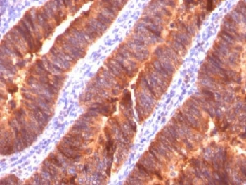
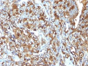
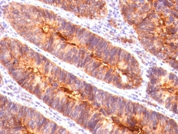
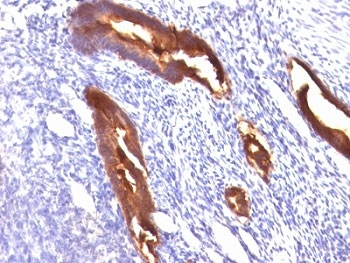
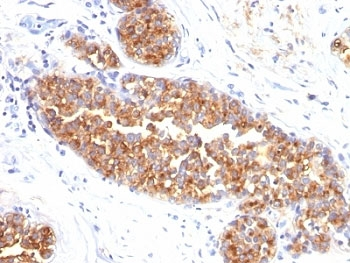
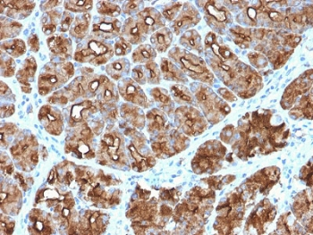
| Description | This mAb recognizes full-length MUC1/Mucin-1/Epithelial Marker Antigen/EMA in a glycosylation-independent manner and can bind to the fully glycosylated protein. The dominant epitope of this mAb is APDTR in the VNTR region. It reacts with the core peptide of the MUC1 protein, which is a member of a family of mucin glycoproteins that are characterized by high carbohydrate content, O-linked oligosaccharides, high molecular weight (>200kDa) and an amino acid composition rich in serine, threonine, proline and glycine. The core protein contains a domain of 20 amino-acid tandem repeats that functions as multiple epitopes for the mAb. Incomplete glycosylation of some tumor-associated mucins may lead to variable unmasking of the multiple peptide epitopes leading to the observed differences in staining intensity between normal and malignant tissues. This mAb reacts with both normal and malignant epithelia of various tissues including breast and colon. |
| Species/Host | Mouse |
| Clonality | Monoclonal |
| Clone Number | HMPV |
| Tested applications | FACS, IF, IHC-P, WB |
| Reactivity | Human |
| Isotype | Mouse IgG1, kappa |
| Immunogen | Human breast cancer cell line ZR-75 cells were used as the immunogen for the MUC-1 antibody. |
| Dilution range | Flow cytometry: 1-2ug/million cells,Immunofluorescence: 1-2ug/ml,Immunohistochemistry (FFPE): 1-2ug/ml for 30 min at RT,Western blot: 1-2ug/ml |
| Purity | Protein G affinity chromatography |
| Conjugation | Unconjugated |
| Formula | 0.2 mg/ml in 1X PBS with 0.1 mg/ml BSA (US sourced) and 0.05% sodium azide |
| Hazard Information | This MUC-1 antibody is available for research use only. |
| UniProt ID | P15941 |
| Storage | Store the MUC-1 antibody at 2-8°C (with azide) or aliquot and store at -20°C or colder (without azide). |
| Buffer/Preservatives | 0.2 mg/ml in 1X PBS with 0.1 mg/ml rAlbumin (US sourced) and 0.05% sodium azide |
| Note | For research use only |
| Application notes | Optimal dilution of the MUC-1 antibody should be determined by the researcher.1. Staining of formalin-fixed tissues requires boiling tissue sections in pH 9 10mM Tris with 1mM EDTA for 10-20 min followed by cooling at RT for 20 min2. The prediluted format is supplied in a dropper bottle and is optimized for use in IHC. After epitope retrieval step (if required), drip mAb solution onto the tissue section and incubate at RT for 30 min. |
| Expiration Date | 12 months from date of receipt. |
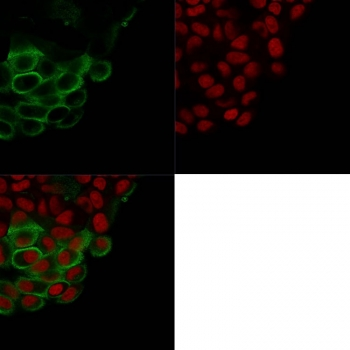
Immunofluorescent staining of PFA-fixed human K562 cells with MUC-1 antibody (green, clone HMPV) and Reddot nuclear stain (red).
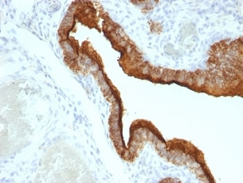
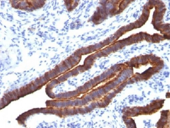
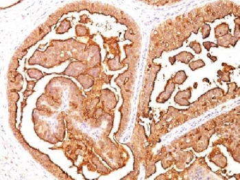
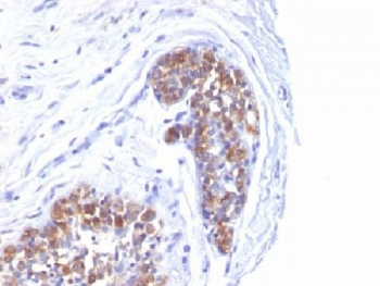
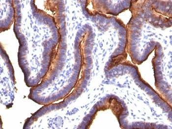
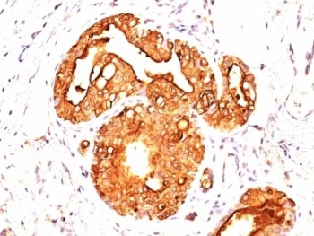
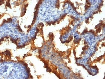
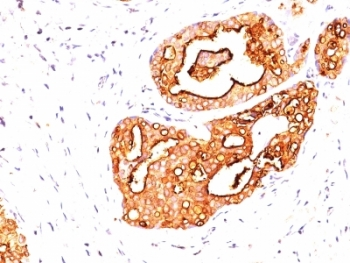
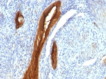
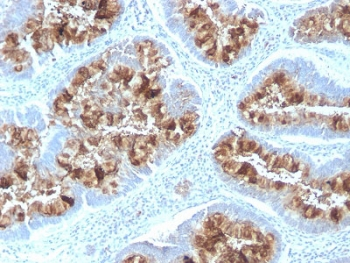
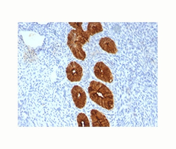
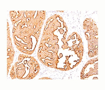
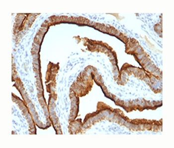
IHC: Formalin-fixed, paraffin-embedded human ovarian carcinoma stained with MUC-1 antibody (clone HMPV).
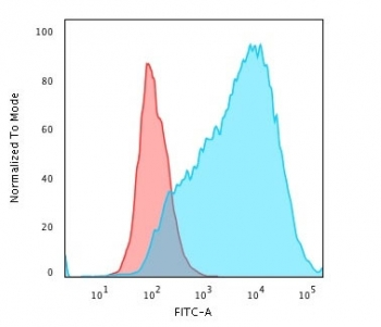
Flow cytometry testing of PFA-fixed human MCF7 cells with MUC-1 antibody (clone HMPV; Red=isotype control, Blue=MUC-1 antibody.
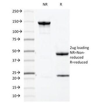
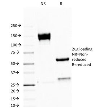
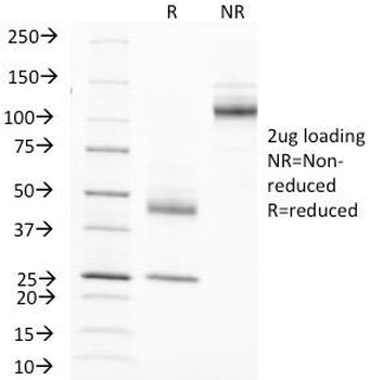
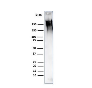
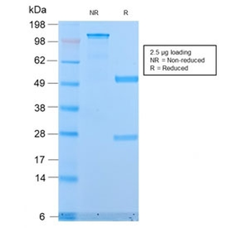
SDS-PAGE analysis of purified, BSA-free MUC-1 antibody (clone HMPV) as confirmation of integrity and purity.
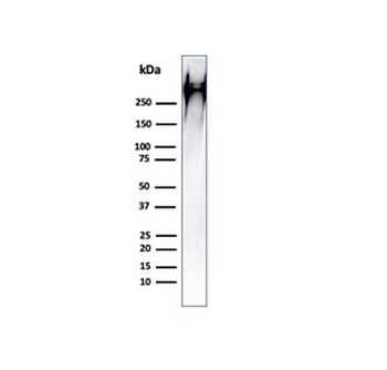
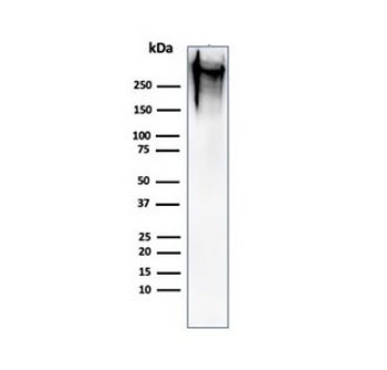
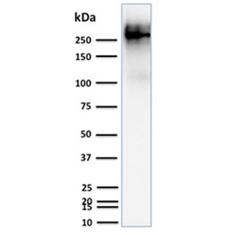
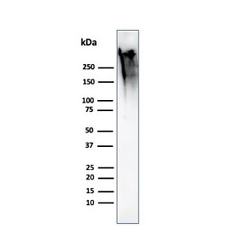
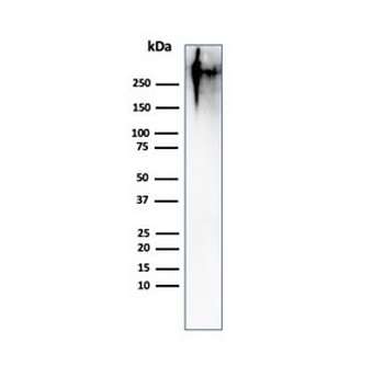
Western blot testing of human MCF7 cell lysate with MUC1 antibody (clone HMPV). This glycoprotein is commonly visualized between 120~500 kDa.
- Item 1 of 8
- Item 1 of 7
- Item 1 of 5
- Item 1 of 4
MUC1 Antibody / Mucin-1 [orb606814]
ELISA, IHC-P, WB
Human
Rabbit
Recombinant
Unconjugated
20 μg, 100 μg - Item 1 of 4

Submit a review
Filter by Rating

- 5 stars

- 4 stars
- 3 stars
- 2 stars
- 1 stars
