You have no items in your shopping cart.
Cart summary
Item 1 of 5
Item 1 of 5
MSRA Antibody (N-term)
Catalog Number: orb1429026

| Catalog Number | orb1429026 |
|---|---|
| Category | Antibodies |
| Description | Rabbit polyclonal antibody to MSRA. |
| Species/Host | Rabbit |
| Clonality | Polyclonal |
| Clone Number | RB18651 |
| Tested applications | FC, IF, IHC-P, WB |
| Reactivity | Human, Mouse |
| Isotype | Rabbit IgG |
| Immunogen | Synthetic Peptide |
| Dilution range | IF: 1:100, WB: 1:1000, WB: 1:1000, IHC-P: 1:10~50, FC: 1:10~50 |
| Form/Appearance | Purified polyclonal antibody supplied in PBS with 0.09% (W/V) sodium azide. This antibody is prepared by Saturated Ammonium Sulfate (SAS) precipitation followed by dialysis against PBS. |
| Conjugation | Unconjugated |
| MW | 26132 |
| Target | MSRA |
| UniProt ID | Q9UJ68 |
| NCBI | NP_001129142.1, NP_001129143.1, NP_001186658.1, NP_036463.1 |
| Storage | Maintain refrigerated at 2-8°C for up to 2 weeks. For long term storage store at -20°C in small aliquots to prevent freeze-thaw cycles |
| Alternative names | Mitochondrial peptide methionine sulfoxide reducta Read more... |
| Note | For research use only |
| Expiration Date | 12 months from date of receipt. |

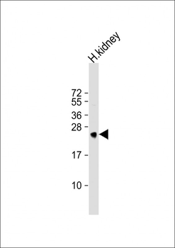
Anti-MSRA Antibody (N-term) at 1:1000 dilution + human kidney lysate.Lysates/proteins at 20 µg per lane. Secondary Goat Anti-Rabbit IgG, (H+L), Peroxidase conjugated at 1/10000 dilution. Predicted band size: 26 kDa. Blocking/Dilution buffer: 5% NFDM/TBST.

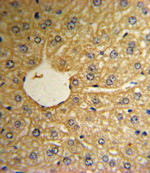
Formalin-fixed and paraffin-embedded human hepatocarcinoma reacted with MSRA Antibody (N-term), which was peroxidase-conjugated to the secondary antibody, followed by DAB staining. This data demonstrates the use of this antibody for immunohistochemistry; clinical relevance has not been evaluated.

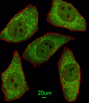
Immunofluorescent analysis of A549 cells, using MSRA Antibody (N-term). Diluted at 1:100 dilution. Alexa Fluor 488-conjugated goat anti-rabbit lgG at 1:400 dilution was used as the secondary antibody (green). Cytoplasmic actin was counterstained with Dylight Fluor 554 (red) conjugated Phalloidin (red).
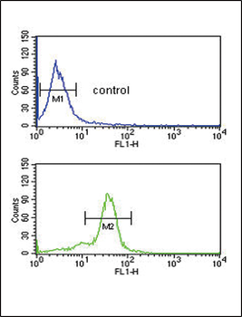
MSRA Antibody (N-term) flow cytometric analysis of MDA-MB435 cells (bottom histogram) compared to a negative control cell (top histogram). FITC-conjugated goat-anti-rabbit secondary antibodies were used for the analysis.

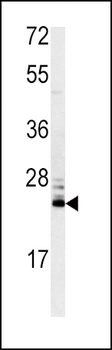
Western blot analysis of MSRA antibody (N-term) in mouse kidney tissue lysates (35 ug/lane). MSRA (arrow) was detected using the purified Pab.
- Item 1 of 5
MSRA Antibody (N-term) [orb1929364]
FC, IF, IHC-P, WB
Human, Mouse
Rabbit
Polyclonal
Unconjugated
400 μl