You have no items in your shopping cart.
Cart summary
Item 1 of 3
Item 1 of 3
Mouse IL-18 antibody
Catalog Number: orb345220

| Catalog Number | orb345220 |
|---|---|
| Category | Antibodies |
| Description | Mouse IL-18 antibody |
| Species/Host | Rabbit |
| Clonality | Polyclonal |
| Tested applications | ELISA, IF, IHC, WB |
| Reactivity | Mouse |
| Isotype | IgG |
| Immunogen | The whole rabbit serum used to produce this IgG fraction antibody was prepared by repeated immunizations with native 157 aa mouse IL-18 produced in E.coli. |
| Concentration | 1.0 mg/mL |
| Dilution range | ELISA: 1:1,000 - 1:5,000, IHC: User Optimized, IF: 1:50-1:200, WB: 1:500 - 1:2,000 |
| Form/Appearance | Liquid (sterile filtered) |
| Purity | This is an IgG preparation of whole rabbit serum purified by protein A chromatography using a low endotoxin methodology. This antibody is primarily directed against mature 18,000 MW mouse IL-18 and is useful in determining its presence in various assays. This antibody will also recognize the 24,000 inactive precursor form of mouse IL-18. In general, this antibody also detects rat IL-18 in the same formats using similar dilutions. A control of similarly diluted LOW ENDOTOXIN CONTROL RABBIT IgG (code # 011-001-297) is recommended. |
| Conjugation | Unconjugated |
| UniProt ID | P70380 |
| NCBI | P70380.2 |
| Storage | Store vial at -20° C prior to opening. Aliquot contents and freeze at -20° C or below for extended storage. Avoid cycles of freezing and thawing. Centrifuge product if not completely clear after standing at room temperature. This product is stable for several weeks at 4° C as an undiluted liquid. Dilute only prior to immediate use. |
| Buffer/Preservatives | None |
| Alternative names | rabbit anti-IL-18 antibody, rabbit anti-interleuki Read more... |
| Note | For research use only |
| Application notes | Anti-Mouse IL-18 has been tested in immunohistochemistry and immunofluorescence and is suitable for use in neutralizations, ELISA, and immunoblotting. Although untested, this reagent may be useful for radioimmunoassays, flow cytometry and immunoprecipitation. It recognizes the 18,000 MW mature (active) IL-18. Reactivity in other immunoassays is unknown. |
| Expiration Date | 12 months from date of receipt. |
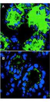
Immunofluorescence microscopy of IL-18 in mouse colon sections. The transversing portion of the large intestine from DSS-exposed (Panel A) and -unexposed mice (Panel B) was excised, rinsed in PBS, and frozen on isopentane cooled with liquid nitrogen. Frozen sections (5 µM) were cut on a Leica CM 1850 cryostat. The slides were fixed for 10 min in 4% paraformaldehyde, air-dried, and incubated for 20 min in PBS supplemented with 10% normal goat serum. Sections were incubated in a 1:50 dilution of Biorbyt's rabbit anti-Mouse IL-18 antibody or 1 µg/ml nonimmune rabbit IgG (not shown) as negative control. The antibodies were diluted in PBS containing 1% bovine serum albumin. After an overnight incubation at 4°C, the sections were washed three times with 0.5% bovine serum albumin in PBS. The sections were then incubated with a secondary goat anti-rabbit antibody conjugated to Alexa488 (Molecular Probes) for 60 min at room temperature in the dark. Nuclei were counterstained blue using 1 µg/100 ml bisbenzimide. After staining, sections were washed and examined with the Leica DM RXA confocal laser scanning system and analyzed. Similar staining will occur with other systems.

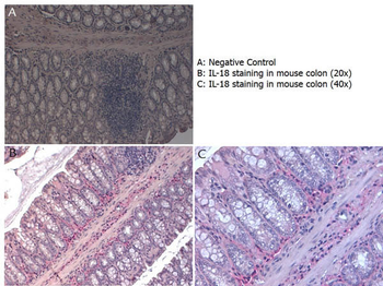
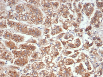
Immunohistochemistry with Rabbit anti-Mouse IL-18 antibody showing IL-18 staining in inflammatory cells of the mucous corium of mouse colon at 20x and 40x. Slide A is a negative control. Slides B and C show staining. Formalin fixed/paraffin embedded sections were subjected to heat induced epitope retrieval (HIER) at pH 6.2 and then incubated with mouse anti-IL-18 antibody at 4.0 µg/ml for 60 minutes. The reaction was developed using MACH 4 universal AP polymer detection system and visualized with WARP RED.

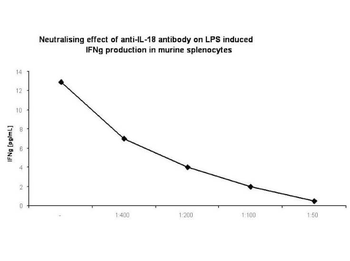
In-vitro neutralization. Spleens were aseptically removed and cell suspensions were prepared. Cells were washed twice and resuspended in RPMI supplemented with 10% FBS. For cytokine measurement, spleen cells were cultured at 5 mln/mL in 24-well, flat-bottom culture plates in the presence of several dilutions of rabbit anti-murine IL-18 antibody (1:400; 1:200; 1:100; 1:50) and 100 ng/mL of LPS (a phenol-extracted preparation from Escherichia coli 055:B5). Cultures were incubated at 37°C in a humidified atmosphere with 5% CO2. At the end of the incubation period, cultures were frozen at -70°C and subjected to 3 freeze-thaw cycles to obtain total cytokine levels. Before assaying, samples were centrifuged for 10 minutes at 10000g to remove debris.
- Item 1 of 3
- Item 1 of 3
- Item 1 of 3
- Item 1 of 3
- Item 1 of 3


Submit a review
Filter by Rating

- 5 stars

- 4 stars
- 3 stars
- 2 stars
- 1 stars
