You have no items in your shopping cart.
Cart summary
Item 1 of 5
Item 1 of 5
Mouse Csf1r Antibody (C-term)
Catalog Number: orb1936300

| Catalog Number | orb1936300 |
|---|---|
| Category | Antibodies |
| Description | Affinity Purified Rabbit Polyclonal Antibody (Pab) |
| Species/Host | Rabbit |
| Clonality | Polyclonal |
| Clone Number | RB34462 |
| Tested applications | IF, IHC-P, WB |
| Reactivity | Mouse, Rat |
| Isotype | Rabbit IgG |
| Antibody Type | Primary Antibody |
| Dilution range | IF: 1:25, WB: 1:1000-2000, WB: 1:1000, WB: 1:1000, IHC-P: 1:25 |
| Form/Appearance | Purified polyclonal antibody supplied in PBS with 0.09% (W/V) sodium azide. This antibody is purified through a protein A column, followed by peptide affinity purification. |
| Conjugation | Unconjugated |
| MW | 109179 Da |
| Target | This Mouse Csf1r antibody is generated from rabbits immunized with a KLH conjugated synthetic peptide between 895-923 amino acids from the C-terminal region of mouse Csf1r. |
| UniProt ID | P09581 |
| NCBI | NP_001032948.2 |
| Storage | Maintain refrigerated at 2-8°C for up to 2 weeks. For long term storage store at -20°C in small aliquots to prevent freeze-thaw cycles |
| Alternative names | Macrophage colony-stimulating factor 1 receptor, C Read more... |
| Note | For research use only |
| Expiration Date | 12 months from date of receipt. |

Mouse Csf1r Antibody (C-term) western blot analysis in mouse heart tissue lysates (35 ug/lane). This demonstrates the Csf1r antibody detected the Csf1r protein (arrow).
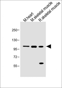
All lanes: Anti-Mouse Csf1r Antibody (C-term) at 1:1000 dilution. Lane 1: Mouse heart lysate. Lane 2: Mouse skeletal muscle lysate. Lane 3: Rat skeletal muscle lysate. Lysates/proteins at 20 µg per lane. Secondary Goat Anti-Rabbit IgG, (H+L), Peroxidase conjugated at 1/10000 dilution. Predicted band size: 109 kDa. Blocking/Dilution buffer: 5% NFDM/TBST.
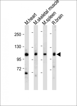
All lanes: Anti-Mouse Csf1r Antibody (C-term) at 1:1000-2000 dilution. Lane 1: Mouse heart tissue lysate. Lane 2: Mouse skeletal muscle tissue lysate. Lane 3: Mouse spleen tissue lysate. Lane 4: Rat brain tissue lysate. Lysates/proteins at 20 µg per lane. Secondary Goat Anti-Rabbit IgG, (H+L), Peroxidase conjugated at 1/10000 dilution. Predicted band size: 109 kDa. Blocking/Dilution buffer: 5% NFDM/TBST.
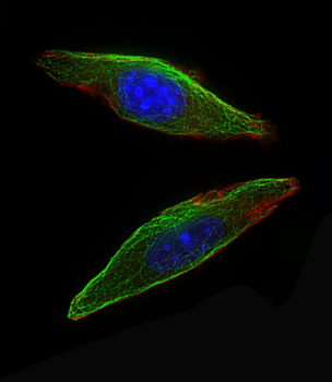
Immunofluorescent analysis of 4% paraformaldehyde-fixed, 0.1% Triton X-100 permeabilized NIH/3T3 cells labeling Csf1r at 1/25 dilution, followed by Dylight 488-conjugated goat anti-Rabbit IgG secondary antibody at 1/200 dilution (green). Immunofluorescence image showing Cytoplasm staining on NIH/3T3 cell line. Cytoplasmic actin is detected with Dylight 554 Phalloidin (red). The nuclear counter stain is DAPI (blue).
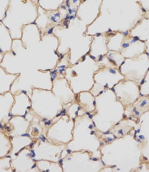
Staining Mouse Csf1r in mouse lung tissue sections by Immunohistochemistry (IHC-P - paraformaldehyde-fixed, paraffin-embedded sections). Tissue was fixed with formaldehyde and blocked with 3% BSA for 0.5 hour at room temperature; antigen retrieval was by heat mediation with a citrate buffer (pH6). Samples were incubated with primary antibody (1/25) for 1 hours at 37°C. A undiluted biotinylated goat polyvalent antibody was used as the secondary antibody.
