You have no items in your shopping cart.
Cart summary
Item 1 of 8
Item 1 of 8
Moesin Antibody
Catalog Number: orb749680

| Catalog Number | orb749680 |
|---|---|
| Category | Antibodies |
| Description | Recognizes 78kDa moesin protein. Moesin, a member of the talin-4.1 superfamily, is a linking protein of the sub-membranous actin cytoskeleton. It is expressed in variable amounts in cells of different phenotypes such as macrophages, lymphocytes, fibroblastic, endothelial, epithelial, and neuronal cell lines but not in blood cells. The ERM proteins, ezrin, radixin, and moesin are involved in a variety of cellular functions, such as cell adhesion, migration, and the organization of cell surface structures, and are highly homologous, both in protein sequence and in functional activity, with merlin/schwannomin, a neurofibromatosis-2-associated tumor-suppressor protein. Cell lines of epithelial and mesothelial origin contain both moesin and radixin whereas cells of endothelial and lymphoid origin express moesin. |
| Species/Host | Mouse |
| Clonality | Monoclonal |
| Clone Number | MSN/493 |
| Tested applications | FACS, IF, IHC-P, WB |
| Reactivity | Human |
| Isotype | Mouse IgG1, kappa |
| Immunogen | Recombinant full-length human protein was used as the immunogen for the Moesin antibody. |
| Dilution range | Flow cytometry: 1-2ug/million cells,Immunofluorescence: 2-4ug/ml,Western blot: 1-2ug/ml,Immunohistochemistry (FFPE): 1-2ug/ml for 30 min at RT |
| Purity | Protein G affinity chromatography |
| Conjugation | Unconjugated |
| Formula | 0.2 mg/ml in 1X PBS with 0.1 mg/ml BSA (US sourced) and 0.05% sodium azide |
| Hazard Information | This Moesin antibody is available for research use only. |
| UniProt ID | P26038 |
| Storage | Store the Moesin antibody at 2-8°C (with azide) or aliquot and store at -20°C or colder (without azide). |
| Buffer/Preservatives | 0.2 mg/ml in 1X PBS with 0.1 mg/ml rAlbumin (US sourced) and 0.05% sodium azide |
| Note | For research use only |
| Application notes | Optimal dilution of the Moesin antibody should be determined by the researcher.1. Staining of formalin-fixed tissues requires boiling tissue sections in 10mM Citrate buffer, pH 6.0, for 10-20 min followed by cooling at RT for 20 minutes2. The prediluted format is supplied in a dropper bottle and is optimized for use in IHC. After epitope retrieval step (if required), drip mAb solution onto the tissue section and incubate at RT for 30 min. |
| Expiration Date | 12 months from date of receipt. |
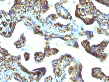
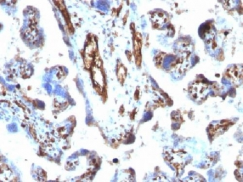
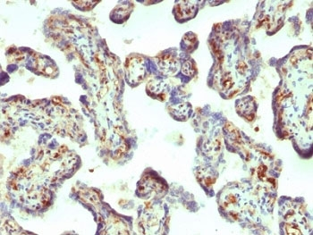
IHC: Formalin-fixed, paraffin-embedded human placenta stained with Moesin antibody (MSN/493). HIER: boil tissue sections in pH 9 10mM Tris with 1mM EDTA for 20 min and allow to cool before testing.
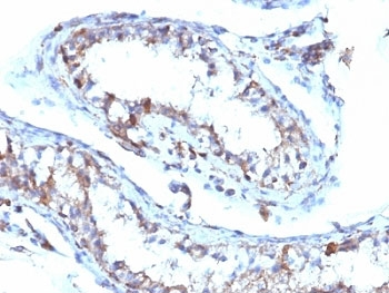
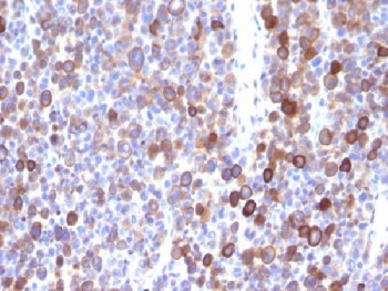
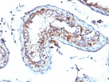
IHC: Formalin-fixed, paraffin-embedded human testicular carcinoma stained with Moesin antibody (MSN/493). HIER: boil tissue sections in pH 9 10mM Tris with 1mM EDTA for 20 min and allow to cool before testing.
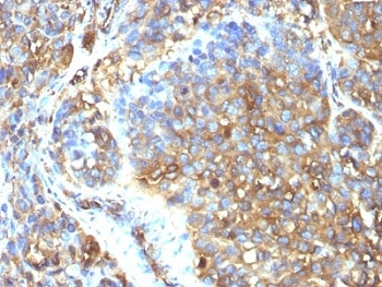
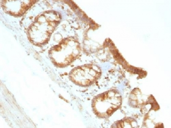
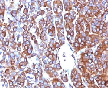
IHC: Formalin-fixed, paraffin-embedded human melanoma stained with Moesin antibody (MSN/493). HIER: boil tissue sections in pH 9 10mM Tris with 1mM EDTA for 20 min and allow to cool before testing.
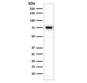
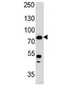
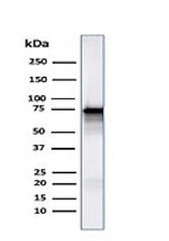
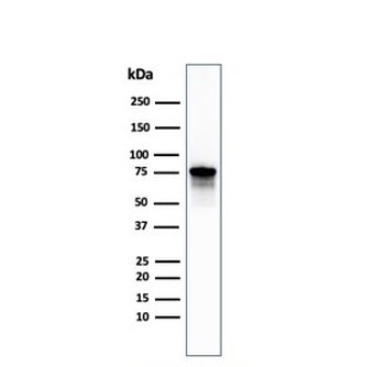
Western blot testing of human Jurkat cell lysate with Moesin antibody. Predicted molecular weight ~68 kDa but routinely observed at 68-78 kDa.
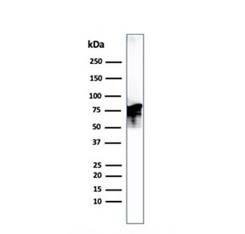
Western blot testing of human PC3 cell lysate with Moesin antibody. Predicted molecular weight ~68 kDa but routinely observed at 68-78 kDa.
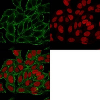
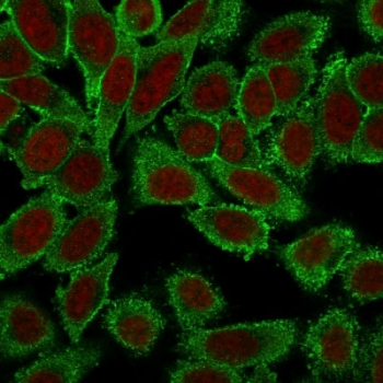
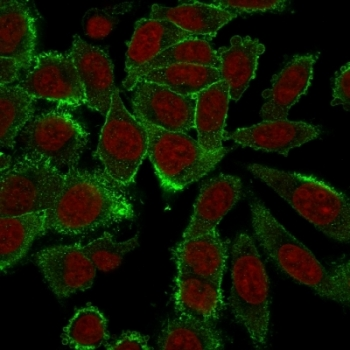
Immunofluorescent staining of PFA-fixed human HeLa cells with Moesin antibody (clone MSN/493, green) and Reddot nuclear stain (red).
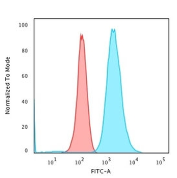
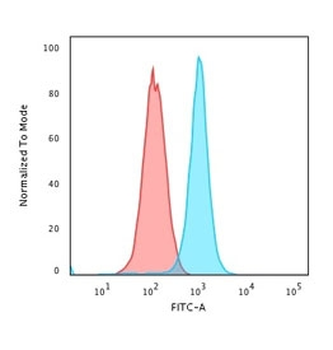
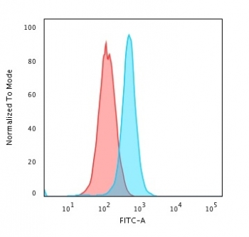
Flow cytometry testing of PFA-fixed human K562 cells with Moesin antibody (clone MSN/493); Red=isotype control, Blue=Moesin antibody.
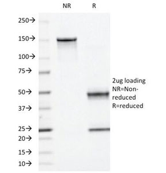
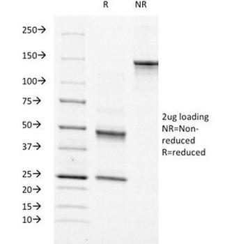
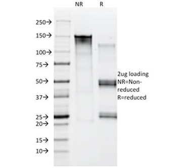
SDS-PAGE Analysis of Purified, BSA-Free Moesin Antibody (clone MSN/493). Confirmation of Integrity and Purity of the Antibody.
- Item 1 of 9
- Item 1 of 8
- Item 1 of 8
NF2/Merlin Antibody [orb570342]
ELISA, FC, ICC, IF, IHC, WB
Human, Mouse, Rat
Rabbit
Polyclonal
Unconjugated
10 μg, 100 μg - Item 1 of 7
Moesin/MSN Antibody (monoclonal, 8D4) [orb865589]
FC, ICC, IF, IHC, WB
Human, Monkey, Mouse, Rat
Mouse
Monoclonal
Unconjugated
10 μg, 100 μg - Item 1 of 6


Submit a review
Filter by Rating

- 5 stars

- 4 stars
- 3 stars
- 2 stars
- 1 stars
