You have no items in your shopping cart.
Cart summary
Item 1 of 13
Item 1 of 13
MMP3 Recombinant Rabbit Monoclonal Antibody
Catalog Number: orb1499363

| Catalog Number | orb1499363 |
|---|---|
| Category | Antibodies |
| Description | MMP3 Recombinant Rabbit Monoclonal Antibody |
| Species/Host | Rabbit |
| Clonality | Recombinant |
| Tested applications | FC, ICC, IF, IHC-Fr, IHC-P, WB |
| Predicted Reactivity | Mouse, Rat |
| Reactivity | Human, Mouse, Rat |
| Isotype | IgG |
| Immunogen | KLH conjugated synthetic peptide derived from human MMP-3 |
| Concentration | 1mg/ml |
| Dilution range | WB=1:500-1000, IHC-P=1:100-500, IHC-F=1:400-800, ICC/IF=1:50, IF=1:50-100, Flow-Cyt=1:100 |
| Form/Appearance | Liquid |
| Conjugation | Unconjugated |
| MW | 54 kDa |
| Target | MMP3 |
| UniProt ID | P08254 |
| Storage | Maintain refrigerated at 2-8°C for up to 2 weeks. For long term storage store at -20°C in small aliquots to prevent freeze-thaw cycles. |
| Buffer/Preservatives | 0.01M TBS (pH7.4) with 1% rAlbumin, 0.02% Proclin300 and 50% Glycerol. |
| Alternative names | MMP3_HUMAN; Stromelysin-1; EC:3.4.24.17; STMY1; SL Read more... |
| Note | For research use only |
| Expiration Date | 12 months from date of receipt. |

Blocking buffer: 5% NFDM/TBST, Primary ab Dilution: 1:1000, Primary ab incubation condition: 2 hours at room temperature, Secondary ab: Goat Anti-Rabbit IgG H&L (HRP), Lysate: 1: Raji, 2: U87-MG, 3: Mouse placenta, 4: Rat placenta, Protein loading quantity: 20 µg, Exposure time: 60 s, Predicted MW: 54 kDa, Observed MW: 54 kDa.
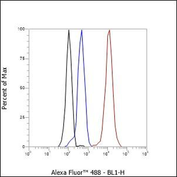
Cell line: HepG2, Fixation: 4% Paraformaldehyde, Permeabilization: 90% Methanol, Primary Ab Dilution: 1:100, Secondary Ab: Goat Anti-Rabbit IgG, Unlabelled control: The cell without incubation with primary antibody and secondary antibody (Black line). Isotype control: Rabbit monoclonal IgG (Blue line). Comment: Line red is the positive signal for orb1499363.
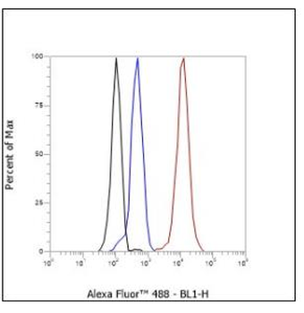
Cell line: HepG2, Fixation: 4% Paraformaldehyde, Permeabilization: 90% Methanol, Primary Ab Dilution: 1:100, Secondary Ab: Goat Anti-Rabbit IgG, Unlabelled control: The cell without incubation, with primary antibody and secondary antibody, (Black line). Isotype control: Rabbit monoclonal IgG (Blue line). Comment: Line red is the positive signal for orb1499363.
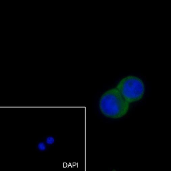
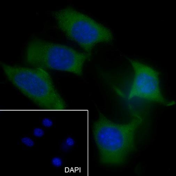
Cell line: HT-29, Fixative: 4% Paraformaldehyde, Permeabilization: 0.1% TritonX-100, Primary ab Dilution: 1:50, Primary incubation condition: 4°C overnight, Secondary ab: Goat Anti-Rabbit IgG, Nuclear counter stain: DAPI (Blue), Comment: Color green is the positive signal for orb1499363.
Cell line: HT-29, Fixative: 4% Paraformaldehyde, Permeabilization: 0.1% TritonX-100, Primary ab Dilution: 1:50, Primary incubation condition: 4°C overnight, Secondary ab: Goat Anti-Rabbit IgG, Nuclear counter stain: DAPI (Blue), Comment: Color green is the positive signal for orb1499363.
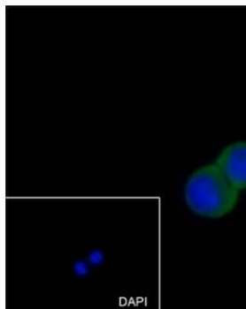
Cell line: NIH/3T3, Fixative: 4% Paraformaldehyde, Permeabilization: 0.1% TritonX-100, Primary ab Dilution: 1:50, Primary incubation condition: 4°C overnight, Secondary ab: Goat Anti-Rabbit IgG, Nuclear counter stain: DAPI (Blue), Comment: Color green is the positive signal for orb1499363.
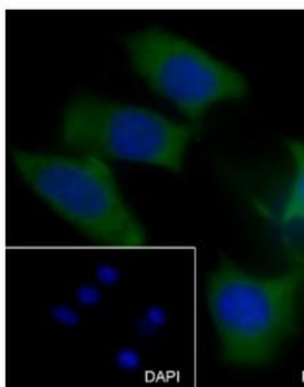
Cell line: NIH/3T3, Fixative: 4% Paraformaldehyde, Permeabilization: 0.1% TritonX-100, Primary ab Dilution: 1:50, Primary incubation condition: 4°C overnight, Secondary ab: Goat Anti-Rabbit IgG, Nuclear counter stain: DAPI (Blue), Comment: Color green is the positive signal for orb1499363.
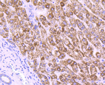
Immunohistochemical analysis of paraffin-embedded human liver tissue using anti-MMP3 antibody. The section was pre-treated using heat mediated antigen retrieval with Tris-EDTA buffer (pH 8.0-8.4) for 20 minutes. The tissues were blocked in 5% BSA for 30 minutes at room temperature, washed with ddH2O and PBS, and then probed with the primary antibody (orb1499363, 1/50) for 30 minutes at room temperature. The detection was performed using an HRP conjugated compact polymer system. DAB was used as the chromogen. Tissues were counterstained with hematoxylin and mounted with DPX.
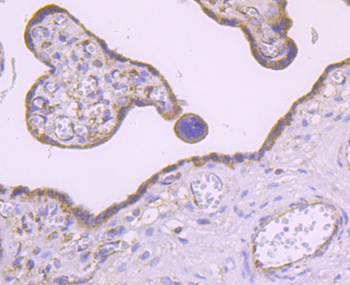
Immunohistochemical analysis of paraffin-embedded human placenta tissue using anti-MMP3 antibody. The section was pre-treated using heat mediated antigen retrieval with Tris-EDTA buffer (pH 8.0-8.4) for 20 minutes. The tissues were blocked in 5% BSA for 30 minutes at room temperature, washed with ddH2O and PBS, and then probed with the primary antibody (orb1499363, 1/50) for 30 minutes at room temperature. The detection was performed using an HRP conjugated compact polymer system. DAB was used as the chromogen. Tissues were counterstained with hematoxylin and mounted with DPX.
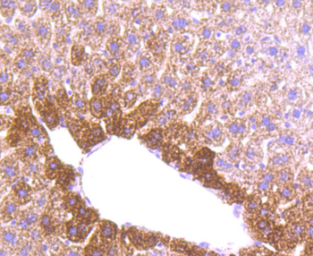
Immunohistochemical analysis of paraffin-embedded mouse liver tissue using anti-MMP3 antibody. The section was pre-treated using heat mediated antigen retrieval with Tris-EDTA buffer (pH 8.0-8.4) for 20 minutes. The tissues were blocked in 5% BSA for 30 minutes at room temperature, washed with ddH2O and PBS, and then probed with the primary antibody (orb1499363, 1/50) for 30 minutes at room temperature. The detection was performed using an HRP conjugated compact polymer system. DAB was used as the chromogen. Tissues were counterstained with hematoxylin and mounted with DPX.
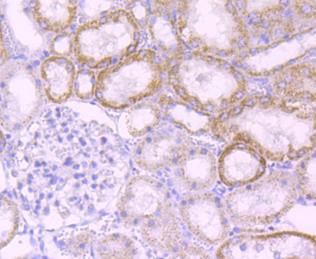
Immunohistochemical analysis of paraffin-embedded rat kidney tissue using anti-MMP3 antibody. The section was pre-treated using heat mediated antigen retrieval with Tris-EDTA buffer (pH 8.0-8.4) for 20 minutes. The tissues were blocked in 5% BSA for 30 minutes at room temperature, washed with ddH2O and PBS, and then probed with the primary antibody (orb1499363, 1/50) for 30 minutes at room temperature. The detection was performed using an HRP conjugated compact polymer system. DAB was used as the chromogen. Tissues were counterstained with hematoxylin and mounted with DPX.
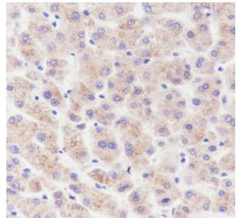
Tissue: Human liver, Section type: Formalin fixed & Paraffin embedded section, Retrieval method: High temperature and high pressure, Retrieval buffer: Tris/EDTA buffer, pH9.0, Primary ab Dilution: 1:100, Primary ab incubation condition: 1 hour at room temperature, Secondary ab: Anti-Rabbit and Mouse, Polymer HRP (Ready to use), Counter stain: Hematoxylin (Blue), Comment: Color brown is the positive signal for orb1499363.
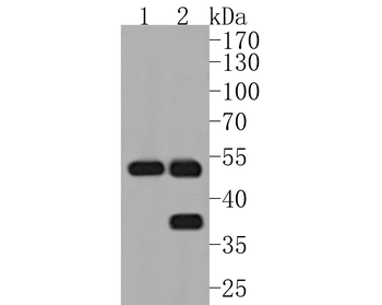
Western blot analysis of MMP3 on different lysates. Proteins were transferred to a PVDF membrane and blocked with 5% BSA in PBS for 1 hour at room temperature. The primary antibody (orb1499363, 1/500) was used in 5% BSA at room temperature for 2 hours. Goat Anti-Rabbit IgG - HRP Secondary Antibody (HA1001) at 1:5000 dilution was used for 1 hour at room temperature. Positive control: Lane 1: human liver tissue lysate, Lane 2: rat liver tissue lysate.

Recombinant MMP3 (Marker of Metastasis and Rheumatoid Arthritis) Antibody [orb534757]
ELISA, FC, WB
Human
Rabbit
Monoclonal
Unconjugated
100 μgMMP3 Recombinant Rabbit Monoclonal Antibody pair (detector) [orb2563253]
ELISA
Rat
Rat
Rabbit
Recombinant
Unconjugated
100 μgMMP3 Recombinant Rabbit Monoclonal Antibody pair (capture) [orb2563254]
ELISA
Rat
Rat
Rabbit
Recombinant
Unconjugated
100 μgMMP3 Recombinant Rabbit Monoclonal Antibody pair (detector) [orb2563255]
ELISA
Human
Human
Rabbit
Recombinant
Unconjugated
100 μgMMP3 Recombinant Rabbit Monoclonal Antibody pair (capture) [orb2563256]
ELISA
Human
Human
Rabbit
Recombinant
Unconjugated
100 μg
