You have no items in your shopping cart.
Cart summary
Item 1 of 3
Item 1 of 3
Mlf1 (phospho-T78) antibody
Catalog Number: orb345549

| Catalog Number | orb345549 |
|---|---|
| Category | Antibodies |
| Description | Mlf1 (phospho-T78) antibody |
| Species/Host | Rabbit |
| Clonality | Polyclonal |
| Tested applications | ELISA, IF, IHC, WB |
| Reactivity | Human |
| Isotype | IgG |
| Immunogen | This affinity purified antibody was prepared from whole rabbit serum produced by repeated immunizations with a synthetic peptide corresponding to amino acids surrounding Thr78 of human MLF1IP protein. The immunogen peptide is phosphorylated at Thr78. |
| Concentration | 1.0 mg/mL |
| Dilution range | ELISA: 1:5,000 - 1:25,000, IHC: 20 µg/ml, IF: User Optimized, WB: 1:500 - 1:2,000 |
| Form/Appearance | Liquid (sterile filtered) |
| Purity | This product was affinity purified from monospecific antiserum by immunoaffinity chromatography using phospho-peptide coupled to agarose beads followed by solid phase adsorption against the non-phospho peptide. This antibody is specific for human MLF1IP protein phosphorylated at Thr78. A BLAST analysis was used to suggest cross-reactivity with MLF1IP protein from human, dog, bovine and chimpanzee based on 100% homology with the immunizing sequence. Expect partial reactivity with homologues from rat and mouse (90% homology). Reactivity against homologues from other sources is not known. |
| Conjugation | Unconjugated |
| UniProt ID | Q71F23 |
| NCBI | 38016935 |
| Storage | Store vial at -20° C prior to opening. Aliquot contents and freeze at -20° C or below for extended storage. Avoid cycles of freezing and thawing. Centrifuge product if not completely clear after standing at room temperature. This product is stable for several weeks at 4° C as an undiluted liquid. Dilute only prior to immediate use. |
| Buffer/Preservatives | 0.01% (w/v) Sodium Azide |
| Alternative names | rabbit anti-MLF1 pT78 antibody, rabbit anti-MLF1 I Read more... |
| Note | For research use only |
| Application notes | This affinity purified antibody has been tested for use in ELISA, western blotting, IF, and IHC. Specific conditions for reactivity should be optimized by the end user. Expect a band approximately 65 kDa in size corresponding to MLF1IP protein by western blotting in the appropriately stimulated tissue, cell lysate or extract. |
| Expiration Date | 12 months from date of receipt. |
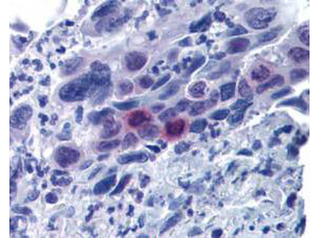
Biorbyt's affinity purified anti-MLF1IP pT78 antibody was used at 20 µg/ml to detect signal in a variety of tissues including multi-human, multi-brain and multi-cancer slides. This image shows moderately positive staining of mitotic cells in colon adenocarcinoma at 60X. Tissue was formalin-fixed and paraffin embedded. The image shows localization of the antibody as the precipitated red signal, with a hematoxylin purple nuclear counterstain.
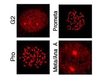
Immunostaining using Biorbyt's affinity purified anti-MLF1IP pT78 antibody shows detection of MLF1IP pT78 at the kinetochores of HeLa cells in different phases of the cell cycle. Fluorescent signals were detectable at the kinetochores as early as G2, became most abundant in prophase cells with a discernible nuclear envelope, and gradually diminished as cells proceeded through mitosis.
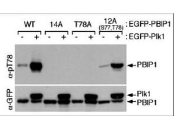
Western blot using Biorbyt's affinity purified anti-MLF1IP pT78 antibody shows detection of MLF1IP phosphorylated at Thr78. HeLa cells were co-infected with the indicated adenoviruses expressing GFP-tagged Plk1 or PBIP1. Blots were probed with the anti-MLF1IP pT78 antibody, stripped, and then reprobed with anti-GFP antibody.

CENPU (phospho-Thr78) Antibody Blocking Peptide [orb1503364]
PBIP1 (phospho-Thr78) antibody [orb100672]
ELISA, IF, IHC-Fr, IHC-P, WB
Bovine, Canine, Equine, Gallus, Human, Mouse, Porcine, Rat, Sheep
Rabbit
Polyclonal
Unconjugated
100 μl, 200 μl, 50 μl
Submit a review
Filter by Rating

- 5 stars

- 4 stars
- 3 stars
- 2 stars
- 1 stars
