You have no items in your shopping cart.
Cart summary
Item 1 of 7
Item 1 of 7
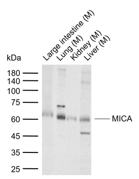
MICA Antibody
Catalog Number: orb1262597

| Catalog Number | orb1262597 |
|---|---|
| Category | Antibodies |
| Description | MICA Antibody |
| Species/Host | Rabbit |
| Clonality | Polyclonal |
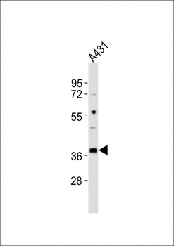
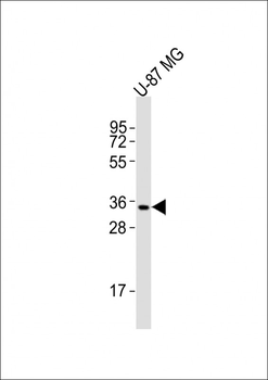
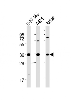
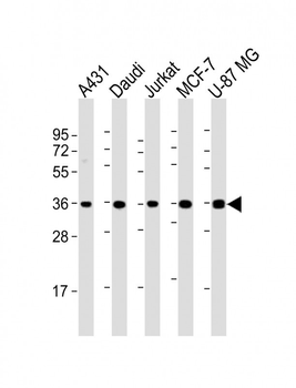
| Tested applications | FC, IF, IHC-P, WB |
| Reactivity | Human |
| Isotype | Rabbit Ig |
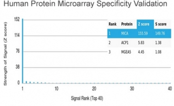
| Immunogen | This MICA antibody is generated from rabbits immunized with a KLH conjugated synthetic peptide between 68-97 amino acids from the Central region of human MICA. |
| Antibody Type | Primary Antibody |
| Concentration | batch dependent |
| Form/Appearance | Liquid |
| Conjugation | Unconjugated |
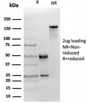
| MW | 43 kDa |
| Target | MICA |
| UniProt ID | Q29983 |
| NCBI | Q29983 |
| Storage | Maintain refrigerated at 2-8°C for up to 2 weeks. For long term storage store at -20°C in small aliquots to prevent freeze-thaw cycles. |
| Buffer/Preservatives | Supplied in PBS with 0.09% (W/V) sodium azide. |
| Alternative names | MHC class I polypeptide-related sequence A, MIC-A, Read more... |
| Note | For research use only |
| Application notes | For IF starting dilution is: 1:25For FACS starting dilution is: 1:25For IHC-P starting dilution is: 1:25 |
| Expiration Date | 12 months from date of receipt. |

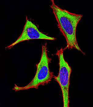
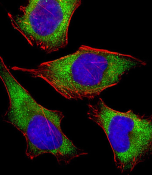
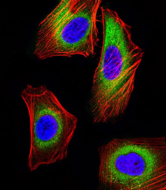
Immunofluorescent analysis of 4% paraformaldehyde-fixed, 0.1% Triton X-100 permeabilized HeLa (human cervical epithelial adenocarcinoma cell line) cells labeling Pdx1 with antibody at 1/25 dilution, followed by 488-conjugated goat anti-rabbit IgG secondary antibody at 1/200 dilution (green). Immunofluorescence image showing cytoplasm staining on HeLa cell line. Cytoplasmic actin is detected with 554 Phalloidin at 1/100 dilution (red). The nuclear counter stain is DAPI (blue).

Immunofluorescent analysis of 4% paraformaldehyde-fixed, 0.1% Triton X-100 permeabilized Hela (Human Cervical epithelial adenocarcinoma cell line) cells labeling Pdx1 with antibody at 1/25 dilution, followed by 488-conjugated goat anti-rabbit IgG secondary antibody at 1/200 dilution (green). Immunofluorescence image showing cytoplasm staining on Hela cell line. Cytoplasmic actin is detected with 554 Phalloidin at 1/100 dilution (red). The nuclear counter stain is DAPI (blue).
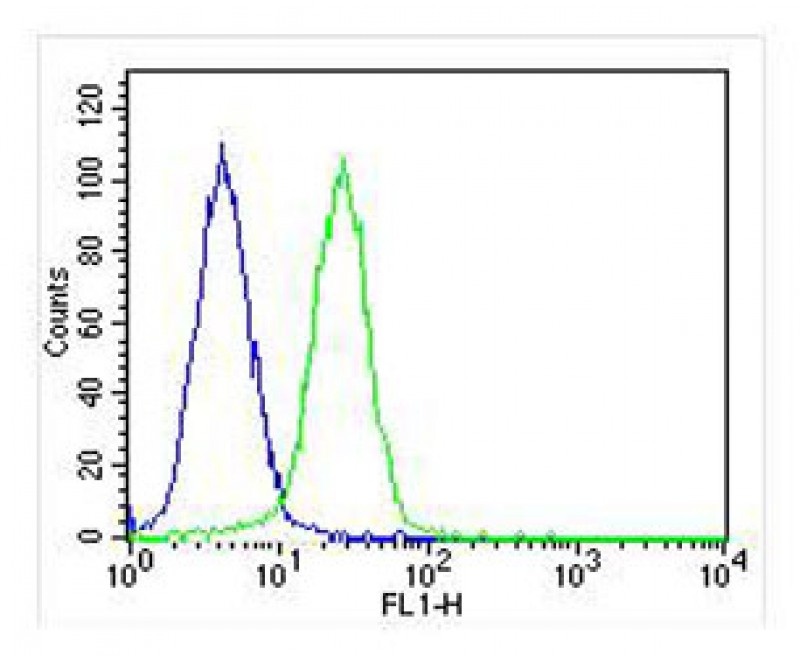
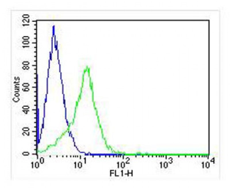
Overlay histogram showing Hela cells stained with Antibody (green line). The cells were fixed with 2% paraformaldehyde (10 min) and then permeabilized with 90% methanol for 10 min. The cells were then icubated in 2% bovine serum albumin to block non-specific protein-protein interactions followed by the antibody (1:25 dilution) for 60 min at 37°C. The secondary antibody used was Goat-Anti-Rabbit IgG, Conjugated Highly Cross-Adsorbed at 1/400 dilution for 40 min at 37°C. Isotype control antibody (blue line) was rabbit IgG (1ug/1x10^6 cells) used under the same conditions. Acquisition of > 10000 events was performed.
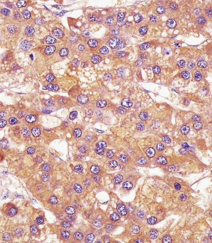
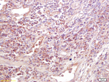
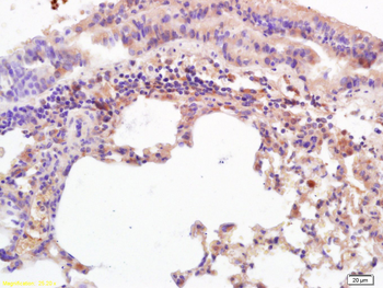
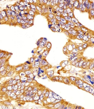
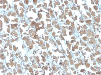
Antibody staining MICA in human hepatic carcinoma tissue sections by Immunohistochemistry (IHC-P - paraformaldehyde-fixed, paraffin-embedded sections).
Immunofluorescent analysis of 4% paraformaldehyde-fixed, 0.1% Triton X-100 permeabilized Hela (Human Cervical epithelial adenocarcinoma cell line) cells labeling Pdx1 with antibody at 1/25 dilution, followed by 488-conjugated goat anti-rabbit IgG secondary antibody at 1/200 dilution (green). Immunofluorescence image showing cytoplasm staining on Hela cell line. Cytoplasmic actin is detected with 554 Phalloidin at 1/100 dilution (red). The nuclear counter stain is DAPI (blue).
Overlay histogram showing SK-BR-3 cells stained with Antibody (green line). The cells were fixed with 2% paraformaldehyde (10 min) and then permeabilized with 90% methanol for 10 min. The cells were then icubated in 2% bovine serum albumin to block non-specific protein-protein interactions followed by the antibody (1:25 dilution) for 60 min at 37°C. The secondary antibody used was Alexa Fluor 488 goat anti-rabbit lgG (H + L) at 1/400 dilution for 40 min at 37°C. Isotype control antibody (blue line) was rabbit IgG1 (1ug/1x10^6 cells) used under the same conditions. Acquisition of > 10000 events was performed.
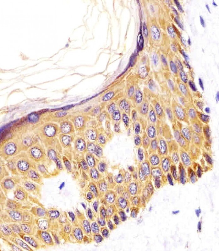
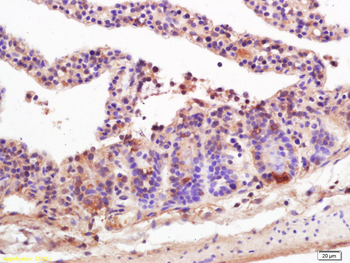
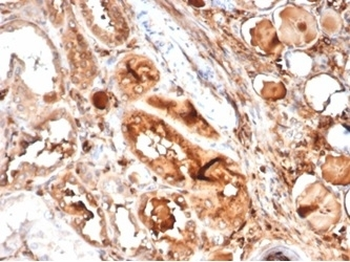
Antibody staining MICA in Human skin tissue sections by Immunohistochemistry (IHC-P - paraformaldehyde-fixed, paraffin-embedded sections).
- Item 1 of 5
MICA Rabbit Polyclonal Antibody [orb11047]
FC, IF, IHC-Fr, IHC-P, WB
Mouse
Human, Mouse
Rabbit
Polyclonal
Unconjugated
50 μl, 100 μl, 200 μl - Item 1 of 7
- Item 1 of 7
MICA Antibody (Center) [orb1928388]
FC, IF, IHC-P, WB
Human
Rabbit
Polyclonal
Unconjugated
50 μl, 100 μl - Item 1 of 4
- Item 1 of 4