You have no items in your shopping cart.
Cart summary
Item 1 of 4
Item 1 of 4
Mesothelin antibody
Catalog Number: orb344443

| Catalog Number | orb344443 |
|---|---|
| Category | Antibodies |
| Description | Mesothelin antibody |
| Species/Host | Mouse |
| Clonality | Monoclonal |
| Clone Number | MB-G10 |
| Tested applications | ELISA, FC, IHC, WB |
| Reactivity | Human |
| Isotype | IgG2a |
| Immunogen | This antibody was produced in mesothelin-deficient mice by immunizations with plasmid cDNA encoding human MSLN full length protein followed by a single boost of a recombinant human mesothelin-Fc fusion protein. |
| Concentration | 1.0 mg/mL |
| Dilution range | ELISA: 1:10,000 - 1:50,000, FC: 1:200, IHC: 1:100, WB: 1:1,000 |
| Form/Appearance | Liquid (sterile filtered) |
| Purity | This antibody is directed against human mesothelin protein. This product was purified from tissue culture supernatant fluid by Protein A chromatography. Cross reactivity with homologues from other sources has not been tested. |
| Conjugation | Unconjugated |
| UniProt ID | Q13421 |
| NCBI | 53988378 |
| Storage | Store vial at -20° C or below prior to opening. This vial contains a relatively low volume of reagent (25 µL). To minimize loss of volume dilute 1:10 by adding 225 µL of the buffer stated above directly to the vial. Recap, mix thoroughly and briefly centrifuge to collect the volume at the bottom of the vial. Use this intermediate dilution when calculating final dilutions as recommended below. Store the vial at -20°C or below after dilution. Avoid cycles of freezing and thawing. |
| Buffer/Preservatives | 0.01% (w/v) Sodium Azide |
| Alternative names | mouse anti-Mesothelin Antibody, Mesothelian, MN, M Read more... |
| Note | For research use only |
| Application notes | This antibody has been tested for use in immunohistochemistry, Flow cytometry, and western blotting. Specific conditions for reactivity should be optimized by the end user. Expect a band approximately 40 kDa in size corresponding to mature mesothelin by western blotting in the appropriate cell lysate or extract. For immunohistochemistry, archival PEFF human tissues were deparaffinized followed by hydration. Antigen-retrieval is recommended. Block tissues with 1% BSA in PBS for 30 min at 23° C. Antibodies are diluted in 1% BSA and reacted with tissue for 60 min at room temperature. |
| Expiration Date | 12 months from date of receipt. |
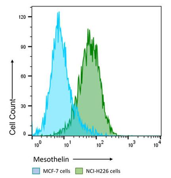
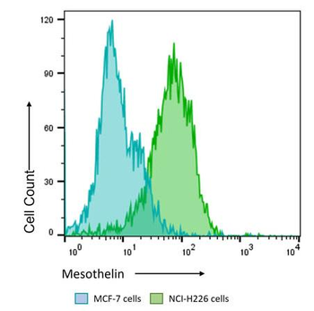
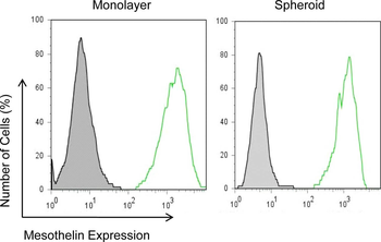
Flow Cytometry Results of Anti-Mesothelin (MOUSE) Monoclonal Antibody. The green histogram shows NCI-H226 cells and blue histogram shows MCF-7 cells. Both cell lines are stained with a 1:200 dilution Anti-Mesothelin (MOUSE) Monoclonal Antibody. The secondary antibody use was Anti-Mouse IgG (H&L) (GOAT) Antibody DyLight™ 488 Conjugated at the 1:400 dilution.
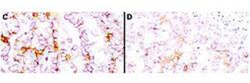
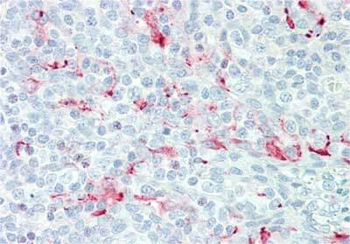
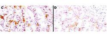
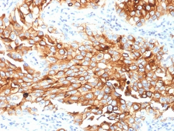
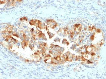
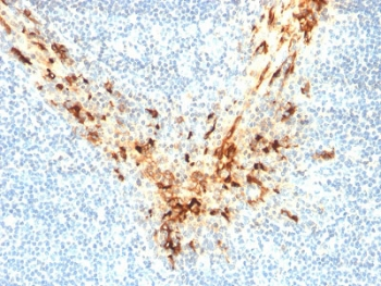
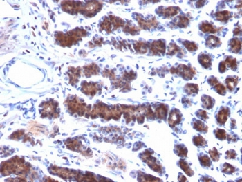
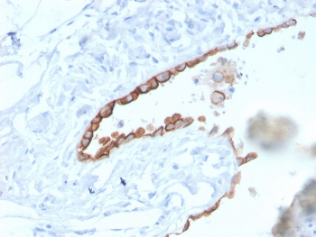
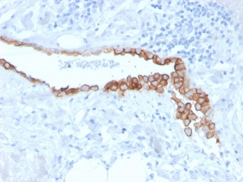
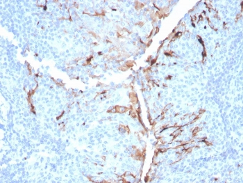
Immunohistochemistry using Biorbyt's anti-mesothelin antibody to react with two epitopes on mesothelin in PEFF human mesothelioma tissue sections treated by antigen retrieval methods. Anti-mesothelin primary antibodies were used at 10 µg/mL to label these sections as follows: C, MAb MB; and D, MAb MN followed by goat anti-mouse IgG conjugated to horseradish peroxidase at 25 µg/mL in 1% BSA/PBS for 30 minutes. (magnification, ×200; bar, 50 μm).
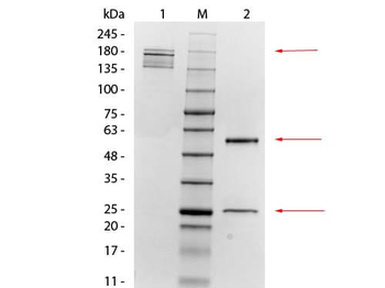
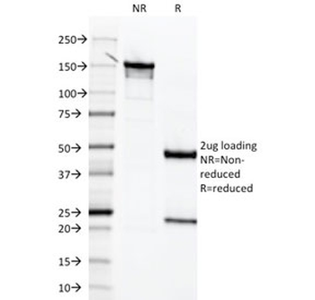
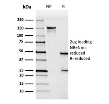
SDS-PAGE of Mouse anti-Mesothelin Monoclonal Antibody. Lane 1: Non-Reduced Mouse anti-Mesothelin Monoclonal Antibody. Lane M: 3 µL OPAL Pre-stained Marker. Lane 2: Reduced Mouse anti-Mesothelin Monoclonal Antibody. Load: 1 µg per lane. Predicted/Observed size: Non-reduced at 160 kDa; Reduced at 55, 25 kDa.
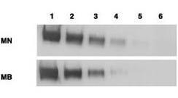
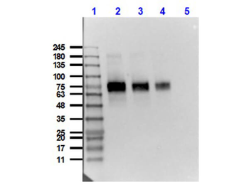
Western blotting using Biorbyt's anti-mesothelin antibody. Load: Mesothelin-Fc (lane 1, 100 ng; lane 2, 25 ng; lane 3, 6 ng; lane 4, 2 ng; and lane 5, 0.4 ng) and CD25-Fc (lane 6, 50 ng) Primary antibody: anti-mesothelin at 1 mg/mL. Secondary Antibody: ALP goat anti-mouse IgG and BCIP/NBT substrate.
- Item 1 of 7
- Item 1 of 7
- Item 1 of 7
Mesothelin Antibody / MSLN [orb606569]
IHC-P
Human, Mouse, Rat
Mouse
Monoclonal
Unconjugated
100 μg, 20 μg - Item 1 of 5
- Item 1 of 5


Submit a review
Filter by Rating

- 5 stars

- 4 stars
- 3 stars
- 2 stars
- 1 stars
