You have no items in your shopping cart.
Cart summary
Item 1 of 4
Item 1 of 4
Mesothelin antibody
Catalog Number: orb344442

| Catalog Number | orb344442 |
|---|---|
| Category | Antibodies |
| Description | Mesothelin antibody |
| Species/Host | Mouse |
| Clonality | Monoclonal |
| Clone Number | MB-G10 |
| Tested applications | ELISA, FC, IHC, WB |
| Reactivity | Human |
| Isotype | IgG2a |
| Immunogen | This antibody was produced in mesothelin-deficient mice by immunizations with plasmid cDNA encoding human MSLN full length protein followed by a single boost of a recombinant human mesothelin-Fc fusion protein. |
| Concentration | 1.0 mg/mL |
| Dilution range | ELISA: 1:10,000 - 1:50,000, FC: 1:200, IHC: 1:100, WB: 1:1,000 |
| Form/Appearance | Liquid (sterile filtered) |
| Purity | This antibody is directed against human mesothelin protein. This product was purified from tissue culture supernatant fluid by Protein A chromatography. Cross reactivity with homologues from other sources has not been tested. |
| Conjugation | Unconjugated |
| UniProt ID | Q13421 |
| NCBI | 53988378 |
| Storage | Store antibody at -20° C prior to opening. Aliquot contents and freeze at -20° C or below for extended storage. Avoid cycles of freezing and thawing. Centrifuge product if not completely clear after standing at room temperature. This product is stable for several weeks at 4° C as an undiluted liquid. Dilute only prior to immediate use. |
| Buffer/Preservatives | 0.01% (w/v) Sodium Azide |
| Alternative names | mouse anti-Mesothelin Antibody, Mesothelian, MN, M Read more... |
| Note | For research use only |
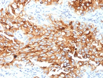
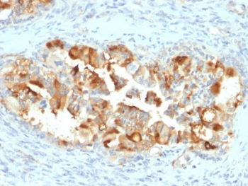
| Application notes | This antibody has been tested for use in immunohistochemistry and western blotting. Specific conditions for reactivity should be optimized by the end user. Expect a band approximately 40 kDa in size corresponding to mature mesothelin by western blotting in the appropriate cell lysate or extract. For Anti-mesothelin immunohistochemistry, archival PEFF human tissues were deparaffinized followed by hydration. Antigen-retrieval is recommended. Block tissues with 1% BSA in PBS for 30 min at 23° C. Antibodies are diluted in 1% BSA and reacted with tissue for 60 min at room temperature. |
| Expiration Date | 12 months from date of receipt. |
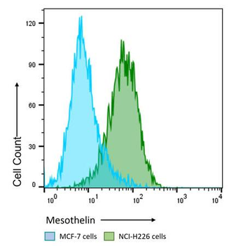
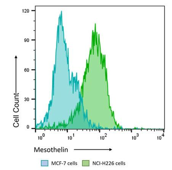
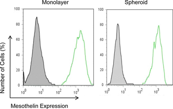
Flow Cytometry Results of Anti-Mesothelin (MOUSE) Monoclonal Antibody. The green histogram shows NCI-H226 cells and blue histogram shows MCF-7 cells. Both cell lines are stained with a 1:200 dilution Anti-Mesothelin (MOUSE) Monoclonal Antibody. The secondary antibody use was Anti-Mouse IgG (H&L) (GOAT) Antibody DyLight™ 488 Conjugated at the 1:400 dilution.
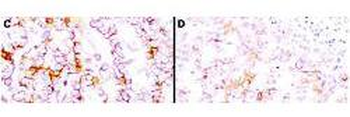
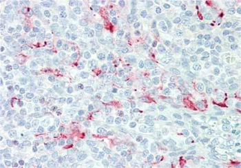
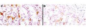
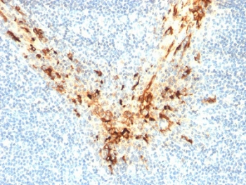
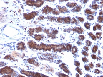
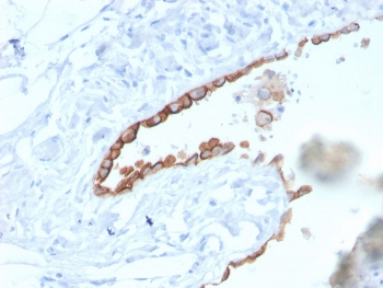
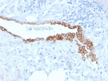
Immunohistochemistry using Biorbyt's anti-mesothelin antibody to react with two epitopes on mesothelin in PEFF human mesothelioma tissue sections treated by antigen retrieval methods. Anti-mesothelin primary antibodies were used at 10 µg/mL to label these sections as follows: C, MAb MB; and D, MAb MN followed by goat anti-mouse IgG conjugated to horseradish peroxidase at 25 µg/mL in 1% BSA/PBS for 30 minutes. (magnification, ×200; bar, 50 μm).
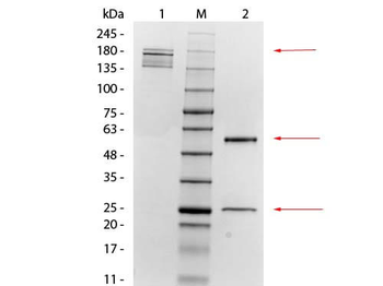
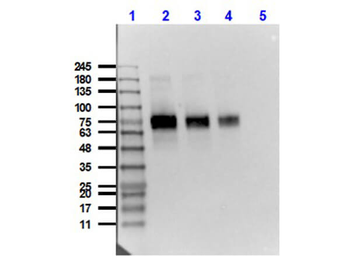
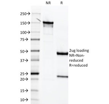
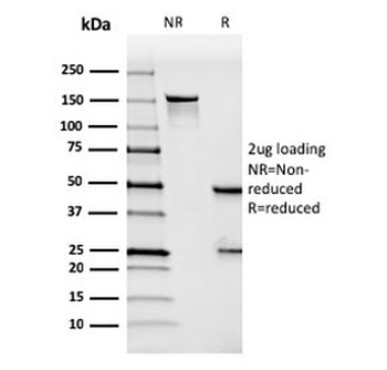
SDS-PAGE of Mouse anti-Mesothelin Monoclonal Antibody. Lane 1: Non-Reduced Mouse anti-Mesothelin Monoclonal Antibody. Lane M: 3 µL OPAL Pre-stained Marker. Lane 2: Reduced Mouse anti-Mesothelin Monoclonal Antibody. Load: 1 µg per lane. Predicted/Observed size: Non-reduced at 160 kDa; Reduced at 55, 25 kDa.
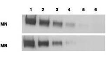
Western blotting using Biorbyt's anti-mesothelin antibody. Load: Mesothelin-Fc (lane 1, 100 ng; lane 2, 25 ng; lane 3, 6 ng; lane 4, 2 ng; and lane 5, 0.4 ng) and CD25-Fc (lane 6, 50 ng) Primary antibody: anti-mesothelin at 1 mg/mL. Secondary Antibody: ALP goat anti-mouse IgG and BCIP/NBT substrate.
- Item 1 of 7
- Item 1 of 7
- Item 1 of 7
Mesothelin Antibody / MSLN [orb606569]
IHC-P
Human, Mouse, Rat
Mouse
Monoclonal
Unconjugated
100 μg, 20 μg - Item 1 of 5
- Item 1 of 5


Submit a review
Filter by Rating

- 5 stars

- 4 stars
- 3 stars
- 2 stars
- 1 stars
