You have no items in your shopping cart.
Cart summary
Item 1 of 4
Item 1 of 4
mCherry antibody
Catalog Number: orb345828

| Catalog Number | orb345828 |
|---|---|
| Category | Antibodies |
| Description | mCherry antibody |
| Species/Host | Rabbit |
| Clonality | Polyclonal |
| Tested applications | ELISA, IF, IHC, WB |
| Reactivity | Other |
| Isotype | IgG |
| Immunogen | The immunogen is a mCherry mutant variant fusion protein of RFP corresponding to the full length amino acid sequence (234aa) derived from the mushroom polyp coral Discosoma. |
| Concentration | 1.04 mg/mL |
| Dilution range | ELISA: 1:150,000 - 1:250,000, IHC: 1:200 - 1:2,000, IF: 1:200 - 1:2,000, WB: 1:2,000 - 1:10,000 |
| Form/Appearance | Liquid (sterile filtered) |
| Purity | mCherry was prepared from monospecific antiserum by immunoaffinity chromatography using Red Fluorescent Protein (Discosoma) coupled to agarose beads followed by solid phase adsorption(s) to remove any unwanted reactivities. Expect reactivity against mCherry, RFP and its variants: tdTomato, mBanana, mOrange, mPlum, mOrange and mStrawberry. Assay by immunoelectrophoresis resulted in a single precipitin arc against anti-Rabbit Serum and purified and partially purified mCherry. No reaction was observed against Human, Mouse or Rat serum proteins. ELISA was used to confirm specificity at less than 0.1% of target signal. |
| Conjugation | Unconjugated |
| Storage | Store vial at -20° C or below prior to opening. This vial contains a relatively low volume of reagent (25 µL). To minimize loss of volume dilute 1:10 by adding 225 µL of the buffer stated above directly to the vial. Recap, mix thoroughly and briefly centrifuge to collect the volume at the bottom of the vial. Use this intermediate dilution when calculating final dilutions as recommended below. Store the vial at -20°C or below after dilution. Avoid cycles of freezing and thawing. |
| Buffer/Preservatives | 0.01% (w/v) Sodium Azide. 0.02 M Potassium Phosphate, 0.15 M Sodium Chloride, pH 7.2 |
| Alternative names | rabbit anti-mCherry antibody, RFP, mCherry monomer Read more... |
| Note | For research use only |
| Application notes | Polyclonal anti-mCherry is designed to detect mCherry, RFP, and its variants. Anti-mCherry (Discosoma sp.) has been tested by ELISA and Western blot and is intended for use in immunological assays including ELISA, western blotting, immunofluorescence, and fluorescence activated cell sorting (FACS). Researchers should determine optimal titers for applications that are not stated. In addition, we performed conjugation of RFP antibodies to either fluorescent dyes, biotin or horseradish peroxidase to further facilitate RFP protein detection and quantification. |
| Expiration Date | 12 months from date of receipt. |
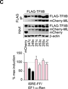
Mapping of a minimal ML sequence required for TFIIB inhibition.A) Schematic representation of ML fragments. B) Top panel: IP of FLAG-tagged TFIIB and GFP-fused ML fragments. Bottom panel: reporter assay in HEK293 cells, where Firefly luciferase under ISRE promoter was co-transfected with EF1-α-Renilla and GFP-ML fragments. C) Top-panel: IP of FLAG-tagged TFIIB and mCherry-fused ML fragments. Bottom panel: reporter assay in HEK293 cells, where Firefly luciferase under ISRE promoter was co-transfected with EF1-α-Renilla and mCherry-ML fragments. Western blots are representative of two experiments with similar results. Bar graphs show mean and SD from three technical replicates and are representative of two experiments with similar results.
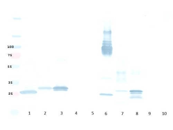
Western Blot of Rabbit Anti-mCherry Antibody MX Hu Ms Rt. Lane 1: Opal Prestained Molecular Weight Marker. Lane 2: RFP (p/n orb345960)/HeLa WCL (p/n orb348668) [0.02 µg/10 µg]. Lane 3: HeLa WCL (p/n orb348668) [10 µg]. Lane 4: RFP (p/n orb345960)/NIH/3T3 WCL (p/n orb348714) [0.02 µg/10 µg]. Lane 5: NIH/3T3 WCL (p/n orb348714) [10 µg]. Lane 6: RFP (p/n orb345960)/PC-12 WCL (p/n orb348733) [0.02 µg/10 µg]. Lane 7: PC-12 WCL (p/n orb348733) [10 µg]. Primary Antibody: Anti-mCherry at 1:1000 overnight at 2-8°C. Secondary Antibody: Goat Anti-Rabbit IgG HRP (p/n orb347654) at 1:70000 for 30 mins at RT. Block: BlockOut Buffer (p/n orb348644). Predicted MW: ~27-30 kDa.

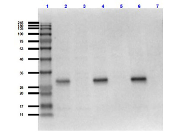
Western Blot of Rabbit anti-mCherry antibody. Lane 1: Biorbyt mCherry fusion protein (reduced). Lane 2: Control dsRed reduced. Lane 3: Control mCherry reduced. Lane 4: Control BFP reduced. Lane 5: Control eGFP reduced. Lane 6: Biorbyt mCherry fusion protein (non-reduced). Lane 7: Control dsRed non-reduced. Lane 8: Control mCherry non-reduced. Lane 9: Control BFP non-reduced. Lane 10: Control eGFP non-reduced. Total load ~200 nanograms per lane. Load: 200 ng per lane. Primary Antibody: Anti-mCherry at 1:5000 for overnight at 4°C. Secondary antibody: HRP rabbit secondary antibody at 1:10000 and orb348656. Block: 5% BLOTTO overnight at 4°C. Predicted/Observed size: 25.9 kDa, for mCherry and RFP, no reaction to BFP or GFP.
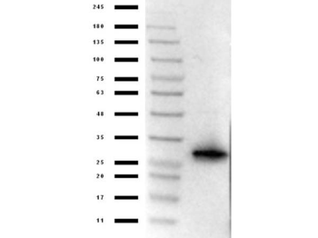
Western Blot of Rabbit Anti-mCherry Antibody. Lane 1: Opal Prestained Marker. Lane 2: 50 ng of RFP. Primary Antibody: rabbit anti-mCherry at 1 µg/mL overnight at 4°C. Secondary Antibody: goat anti-Rabbit peroxidase (p/n orb347654) at 1:70000 for 30 mins at RT. Block: BlockOut Universal Buffer (p/n orb348644). Expect band ~30 kDa.
- Item 1 of 2
- Item 1 of 3
- Item 1 of 2
- Item 1 of 2
- Item 1 of 4


Submit a review
Filter by Rating

- 5 stars

- 4 stars
- 3 stars
- 2 stars
- 1 stars
