You have no items in your shopping cart.
Cart summary
Item 1 of 2
Item 1 of 2
Mast Cell Chymase/CMA1 Antibody
Catalog Number: orb182387

| Catalog Number | orb182387 |
|---|---|
| Category | Antibodies |
| Description | Mast Cell Chymase/CMA1 Antibody |
| Species/Host | Rabbit |
| Clonality | Polyclonal |
| Tested applications | IHC, WB |
| Reactivity | Human, Mouse, Rat |
| Isotype | Rabbit IgG |
| Immunogen | E.coli-derived human CMA1 recombinant protein (Position: I22-N247). Human CMA1 shares 75% and 74% amino acid (aa) sequences identity with mouse and rat CMA1, respectively. |
| Concentration | Adding 0.2 ml of distilled water will yield a concentration of 500 μg/ml. |
| Form/Appearance | Lyophilized |
| Conjugation | Unconjugated |
| MW | 27325 MW |
| UniProt ID | P23946 |
| Storage | Store at -20˚C for one year from date of receipt. After reconstitution, at 4˚C for one month. It can also be aliquotted and stored frozen at -20˚C for six months. Avoid repeated freeze-thaw cycles. |
| Alternative names | Chymase;3.4.21.39;Alpha-chymase;Mast cell protease Read more... |
| Note | For research use only |
| Application notes | WB: The detection limit for CMA1 is approximately 0.25ng/lane under reducing conditions. Tested Species: In-house tested species with positive results. By Heat: Boiling the paraffin sections in 10mM citrate buffer, pH6.0, for 20mins is required for the staining of formalin/paraffin sections. Other applications have not been tested. Optimal dilutions should be determined by end users. . Add 0.2ml of distilled water will yield a concentration of 500ug/ml. |
| Expiration Date | 12 months from date of receipt. |
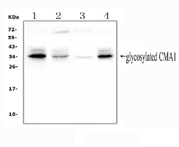
WB analysis of CMA1 using anti-CMA1 antibody.Lane 1:human PC-3 cell;2:human HepG2 cell;3:rat liver tissue;4:mouse HEPA1-6 cell.
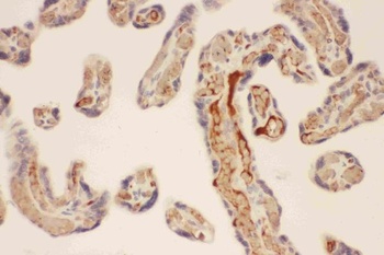
IHC analysis of CMA1 using anti-CMA1 antibody. CMA1 was detected in a paraffin-embedded section of human placenta tissue.

Mast Cell Chymase (CMA1) polyclonal antibody [orb1787180]
WB
Human, Mouse
Rabbit
Polyclonal
Unconjugated
200 μg, 100 μg, 50 μgMast Cell Chymase Rabbit mAb Antibody [orb1924624]
IHC-P, WB
Human
Rabbit
Monoclonal
Unconjugated
100 μl, 50 μl, 20 μl
Submit a review
Filter by Rating

- 5 stars

- 4 stars
- 3 stars
- 2 stars
- 1 stars
