You have no items in your shopping cart.
Cart summary
Item 1 of 6
Item 1 of 6
MANF Antibody
Catalog Number: orb1239620

| Catalog Number | orb1239620 |
|---|---|
| Category | Antibodies |
| Description | MANF Antibody |
| Species/Host | Rabbit |
| Clonality | Polyclonal |
| Tested applications | ELISA, IF, IHC-P, WB |
| Predicted Reactivity | Bovine |
| Reactivity | Human, Mouse, Rat |
| Isotype | IgG |
| Immunogen | Anti-MANF antibody (orb1239620) was raised against a peptide corresponding to 12 amino acids near the amino terminus of human MANF. The immunogen is located within the first 50 amino acids of MANF. |
| Concentration | 1 mg/mL |
| Form/Appearance | Liquid |
| Conjugation | Unconjugated |
| MW | Predicted: 20kDObserved: 26 kD |
| Target | MANF |
| UniProt ID | P55145 |
| NCBI | P55145 |
| Storage | Maintain refrigerated at 2-8°C for up to 2 weeks. For long term storage store at -20°C in small aliquots to prevent freeze-thaw cycles. |
| Buffer/Preservatives | MANF Antibody is supplied in PBS containing 0.02% sodium azide. |
| Alternative names | MANF Antibody: ARP, ARMET, ARP, Mesencephalic astr Read more... |
| Note | For research use only |
| Expiration Date | 12 months from date of receipt. |

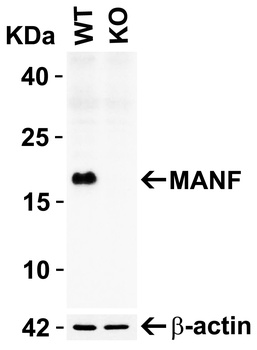
Western Blot Validation in Human Cell Lines. Loading: 15 µg of lysates per lane. Antibodies: MANF orb1239620, (2 µg/mL), 1h incubation at RT in 5% NFDM/TBST. Secondary: Goat anti-rabbit IgG HRP conjugate at 1:10000 dilution.

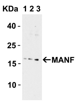
Western Blot Validation with Recombinant Protein. Loading: 30 ng of human MANF recombinant protein per lane. Antibodies: MANF orb1239620 (Lane 1: 0.125 µg/mL, Lane 2: 0.25 µg/mL and Lane 3: 0.5 µg/mL), 1h incubation at RT in 5% NFDM/TBST. Secondary: Goat anti-rabbit IgG HRP conjugate at 1:10000 dilution. Observed at around 55kD.

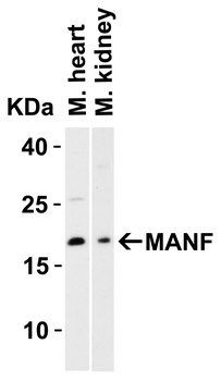
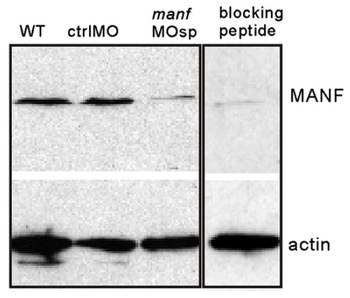
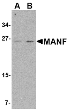
Western Blot Validation in Rat Brain Tissue Lysate. Loading: 15 µg of lysates per lane. Antibodies: MANF orb1239620 (A: 1 µg /mL and B: 2 µg /mL), 1h incubation at RT in 5% NFDM/TBST. Secondary: Goat anti-rabbit IgG HRP conjugate at 1:10000 dilution.

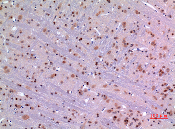
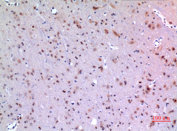
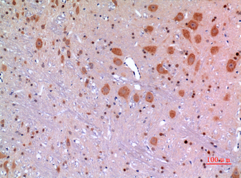
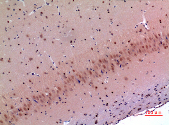
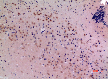
Immunohistochemistry Validation of MANF in Human Brain Tissue. Immunohistochemical analysis of paraffin-embedded human brain tissue using anti-MANF antibody (orb1239620) at 2.5 µg /ml. Tissue was fixed with formaldehyde and blocked with 10% serum for 1 h at RT; antigen retrieval was by heat mediation with a citrate buffer (pH6). Samples were incubated with primary antibody overnight at 4°C. A goat anti-rabbit IgG H&L (HRP) at 1/250 was used as secondary. Counter stained with Hematoxylin.

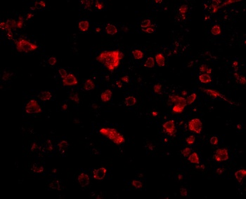
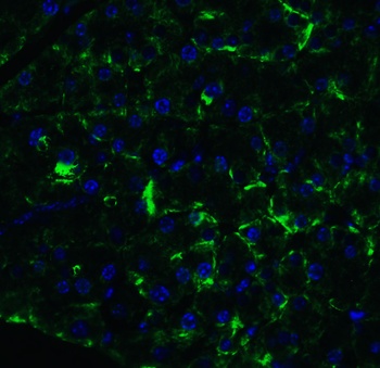
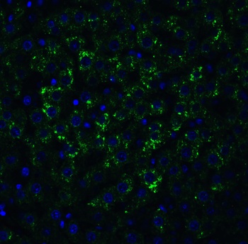
Immunofluorescence Validation of MANF in Human Brain Tissue. Immunofluorescent analysis of 4% paraformaldehyde-fixed human brain tissue labeling MANF with orb1239620 at 20 µg /mL, followed by goat anti-rabbit IgG secondary antibody at 1/500 dilution (red).

Induced Expression Validation of MANF in Mouse C17.2 Neural Stem Cells (Almutawaa et al., 2014). Concentration- and time-dependent effects of Valproic acid (VPA) on MANF at 48 h examined by Western blot analysis with anti-MANF antibodies. MANF was markedly increased 48h after VPA treatment. Lanes 1–4: control, 0.5 mM VPA, 1 mM VPA, 3 mM VPA.
- Item 1 of 15
MANF Antibody [orb1239622]
ELISA, IF, IHC-P, WB
Bovine
Human, Mouse, Rat
Rabbit
Polyclonal
Unconjugated
0.1 mg - Item 1 of 6
- Item 1 of 4
- Item 1 of 2
MANF/ARMET Antibody [orb1537855]
ELISA, IF, IHC, IHC-P, WB
Human, Mouse, Rat
Rabbit
Polyclonal
Unconjugated
0.05 mg - Item 1 of 2