You have no items in your shopping cart.
Cart summary
Item 1 of 4
Item 1 of 4
LSD1 Antibody (N-term)
Catalog Number: orb1937675

| Catalog Number | orb1937675 |
|---|---|
| Category | Antibodies |
| Description | Purified Rabbit Polyclonal Antibody (Pab) |
| Species/Host | Rabbit |
| Clonality | Polyclonal |
| Clone Number | RB7579; RB7580 |
| Tested applications | IF, IHC-P, WB |
| Reactivity | Human, Mouse |
| Isotype | Rabbit IgG |
| Antibody Type | Primary Antibody |
| Dilution range | IF: 1:10~50, WB: 1:1000, WB: 1:1000, IHC-P: 1:50~100 |
| Form/Appearance | Purified polyclonal antibody supplied in PBS with 0.09% (W/V) sodium azide. This antibody is prepared by Saturated Ammonium Sulfate (SAS) precipitation followed by dialysis against PBS. |
| Conjugation | Unconjugated |
| MW | 92903 Da |
| Target | This LSD1 antibody is generated from rabbits immunized with a KLH conjugated synthetic peptide between 108-142 amino acids from the N-terminal region of human LSD1. |
| UniProt ID | O60341 |
| NCBI | NP_001009999.1, NP_055828.2 |
| Storage | Maintain refrigerated at 2-8°C for up to 2 weeks. For long term storage store at -20°C in small aliquots to prevent freeze-thaw cycles |
| Alternative names | Lysine-specific histone demethylase 1A, 1---, BRAF Read more... |
| Note | For research use only |
| Expiration Date | 12 months from date of receipt. |

Fluorescent confocal image of Hela cell stained with LSD1 Antibody (N-term). Hela cells were fixed with 4% PFA (20 min), permeabilized with Triton X-100 (0.1%, 10 min), then incubated with LSD1 primary antibody (1:25, 1 h at 37°C). For secondary antibody, Alexa Fluor 488 conjugated donkey anti-rabbit antibody (green) was used (1:400, 50 min at 37°C). Cytoplasmic actin was counterstained with Alexa Fluor 555 (red) conjugated Phalloidin (7 units/ml, 1 h at 37°C). Nuclei were counterstained with DAPI (blue) (10 µg/ml, 10 min). LSD1 immunoreactivity is localized to Cytoplasm significantly and Nucleus weakly.
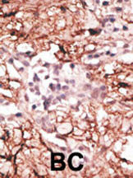
Formalin-fixed and paraffin-embedded human cancer tissue reacted with the primary antibody, which was peroxidase-conjugated to the secondary antibody, followed by DAB staining. This data demonstrates the use of this antibody for immunohistochemistry; clinical relevance has not been evaluated. BC = breast carcinoma; HC = hepatocarcinoma.
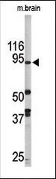
Western blot analysis of anti-LSD1 Pab in mouse brain tissue lysate (35 ug/lane). LSD1 (arrow) was detected using the purified Pab.
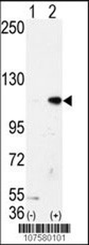
Western blot analysis of AOF2 (arrow) using LSD1 Antibody (N-term).293 cell lysates (2 ug/lane) either nontransfected (Lane 1) or transiently transfected with the AOF2 gene (Lane 2).
