You have no items in your shopping cart.
Cart summary
Item 1 of 16
Item 1 of 16
LHCGR antibody
Catalog Number: orb13542

| Catalog Number | orb13542 |
|---|---|
| Category | Antibodies |
| Description | LHCGR antibody |
| Species/Host | Rabbit |
| Clonality | Polyclonal |
| Tested applications | ICC, IF, IHC-P, WB |
| Reactivity | Human, Mouse, Porcine, Rat |
| Isotype | IgG |
| Immunogen | KLH conjugated synthetic peptide derived from human LHCGR. Please contact us for the exact immunogen sequence. The peptide is available as orb12912. |
| Concentration | - 100 μg (in 200 μl): 0.5 mg/ml- 200 μg (in 400 μl): 0.5 mg/ml |
| Dilution range | IF/ICC: 1:50-400, WB: 1:200-1000, IHC-P: 1:50-400 |
| Form/Appearance | 10 mM PBS, 0.02% sodium azide |
| Purity | Polyclonal antibodies are purified by peptide affinity chromatography |
| Conjugation | Unconjugated |
| MW | 78 kDa |
| Target | LHCGR |
| Entrez | 16867 |
| UniProt ID | P16235, P30730 |
| NCBI | 013582, 11, 038610, 21 |
| RRID | AB_10750213 |
| Storage | Maintain refrigerated at 2-8°C for up to 2 weeks. For long term storage store at -20°C in small aliquots to prevent freeze-thaw cycles. |
| Alternative names | anti-Gonadotropin receptor antibody, anti-GTHR-II Read more... |
| Note | For research use only |
| Expiration Date | 12 months from date of receipt. |
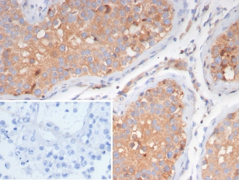
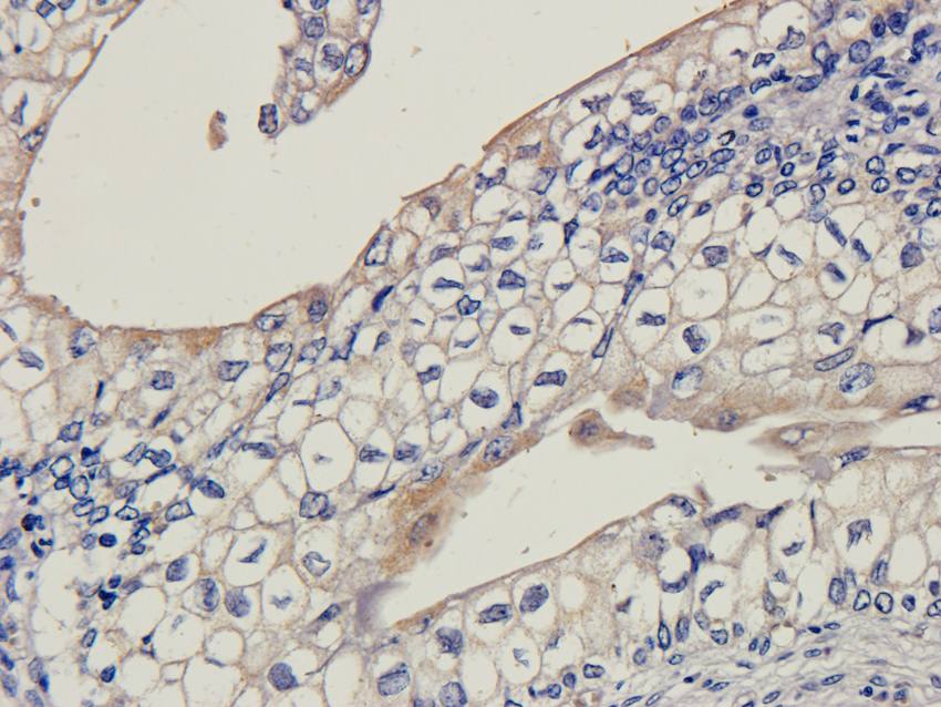
Immunohistochemical staining of mouse skin tissue using LHCGR antibody (dilution of primary antibody - 1:100)

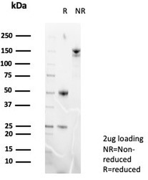
WB analysis of rat thyroid (lane 1), rat epididymis (lane 2), mouse ovary (lane 3), mouse testis (lane 4), mouse uterus (lane 5), mouse brain (lane 6) using LHCGR antibody (1 ug/ml)
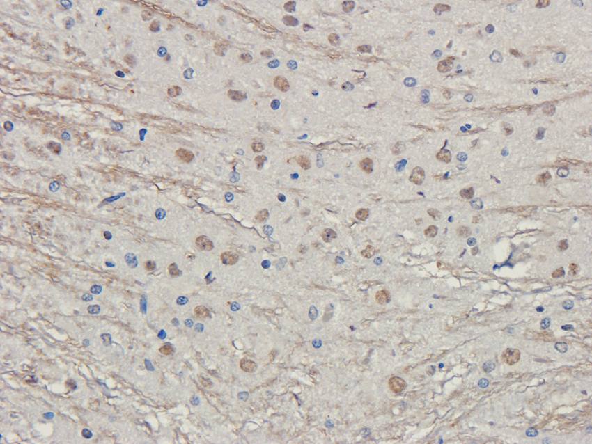
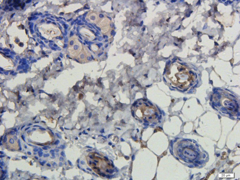
IHC-P image of rat brain tissue using LHCG Receptor antibody (2.5 ug/ml)
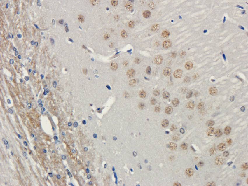
IHC-P image of rat brain tissue using LHCG Receptor antibody (2.5 ug/ml)
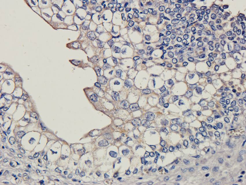
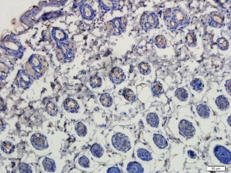
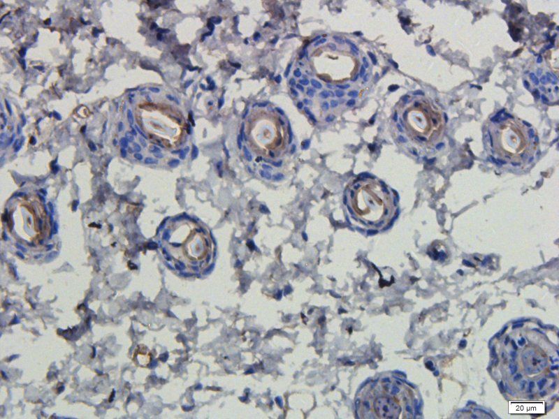
IHC-P image of pig uterus tissue using anti-LHCG Receptor (2.5 ug/ml)
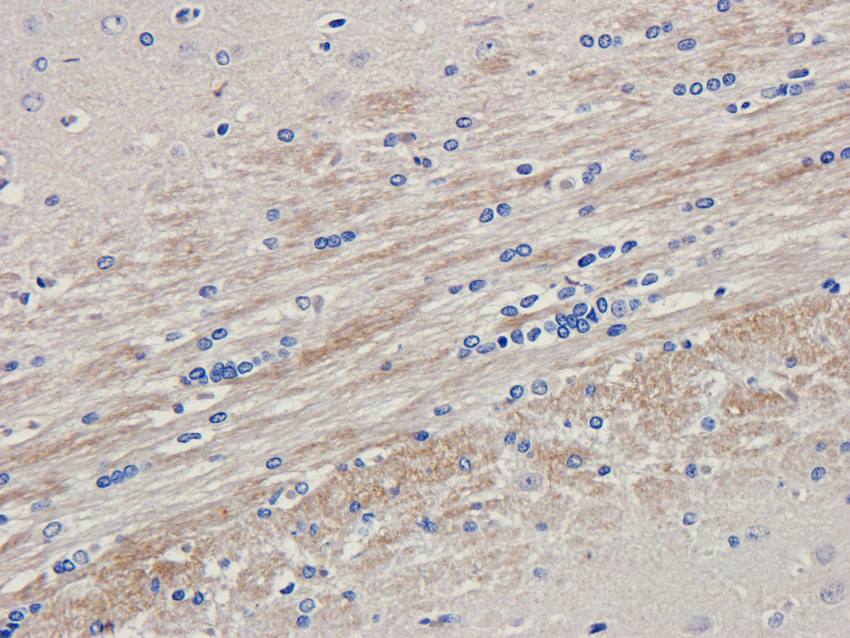
Immunohistochemical staining of rat brain tissue using anti-LHCG Receptor (2.5 ug/ml)
Immunohistochemical staining of pig uterus tissue using LHCG Receptor antibody (2.5 ug/ml)
Immunofluorescence image of mouse skin tissue using LHCGR antibody (dilution at 1:100)
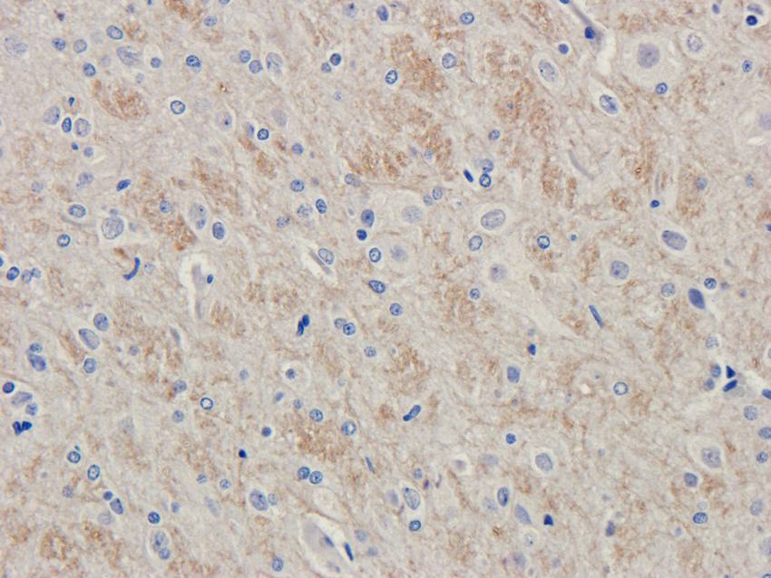
IHC-P staining of rat brain tissue using LHCG Receptor antibody (2.5 ug/ml)
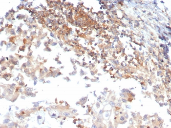
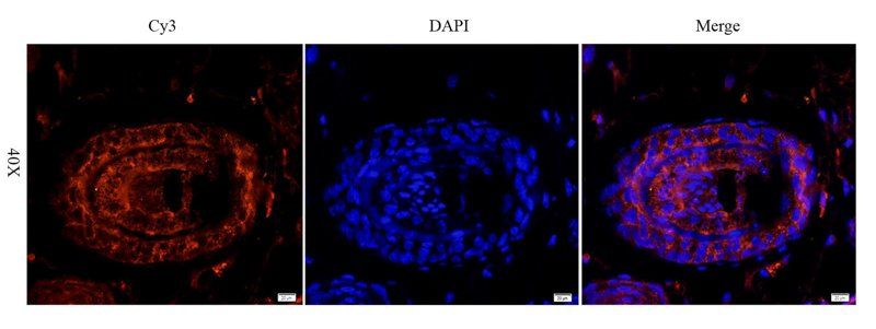
Immunofluorescence analysis of pig large intestines tissue using anti-LHCGR (dilution of primary antibody - 1:100)
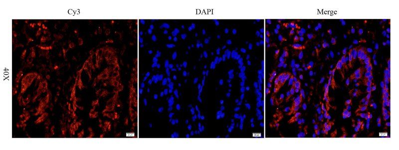
IF analysis of pig large intestines tissue using LHCGR antibody (dilution of primary antibody at 1:100)
IHC-P image of mouse skin tissue using LHCGR antibody (dilution of primary antibody at 1:100)
IHC-P staining of mouse skin tissue using anti-LHCGR (dilution at 1:100)
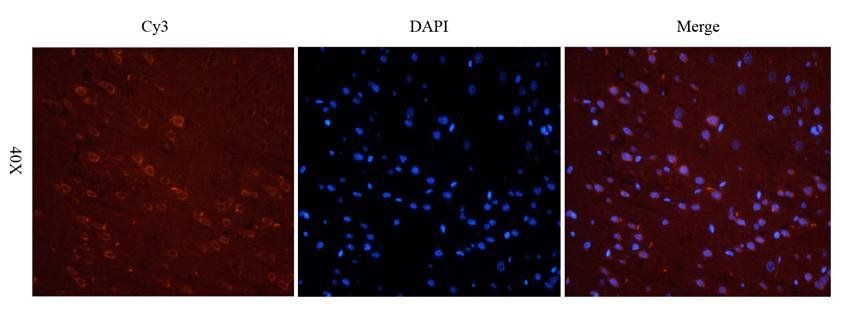
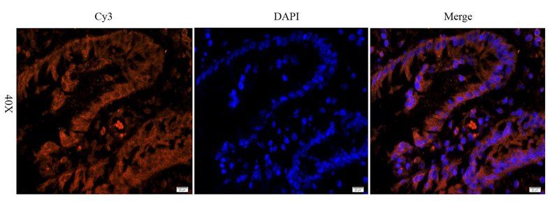
Immunofluorescence image of rat brain tissue using LHCG Receptor antibody (2.5 ug/ml)
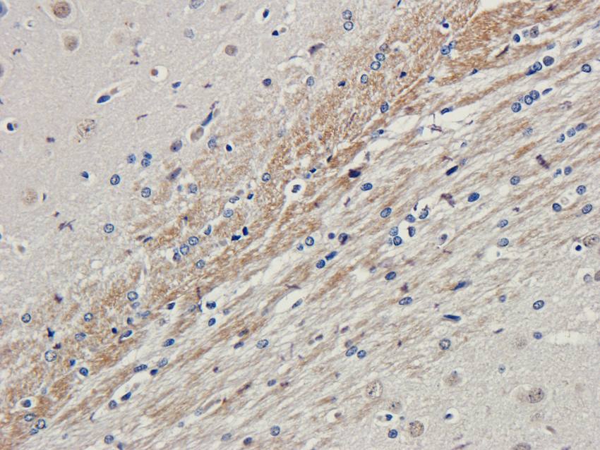
IHC-P staining of rat brain tissue using LHCG Receptor antibody (2.5 ug/ml)
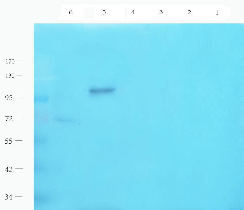
Western blot analysis of rat thyroid (lane 1), rat epididymis (lane 2), mouse ovary (lane 3), mouse testis (lane 4), mouse uterus (lane 5), mouse brain (lane 6) using LHCGR antibody (1 ug/ml)
- Item 1 of 4
- Item 1 of 4
- Item 1 of 3
- Item 1 of 3
- Item 1 of 3