You have no items in your shopping cart.
Cart summary
Item 1 of 15
Item 1 of 15

| Catalog Number | orb33328 |
|---|---|
| Category | Antibodies |
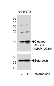
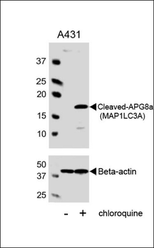
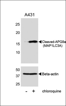
| Description | LC3 antibody |
| Species/Host | Rabbit |
| Clonality | Polyclonal |
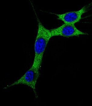
| Tested applications | ICC, IF, IHC-P, WB |
| Predicted Reactivity | Bovine |
| Reactivity | Human, Mouse, Rat |
| Isotype | IgG |
| Immunogen | KLH conjugated synthetic peptide derived from human LC3. Please contact us for the exact immunogen sequence. The peptide is available as orb12928. |
| Concentration | - 100 μg (in 200 μl): 0.5 mg/ml- 200 μg (in 400 μl): 0.5 mg/ml |
| Dilution range | IF/ICC: 1:100-800, IHC-P: 1:100-800, WB: 1:200-2000 |
| Form/Appearance | 10 mM PBS, 0.02% sodium azide |
| Purity | Polyclonal antibodies are purified by peptide affinity chromatography |
| Conjugation | Unconjugated |
| MW | 14 kDa |
| Target | LC3 |
| Entrez | 81631 |
| UniProt ID | Q9GZQ8 |
| RRID | AB_10995488 |
| Storage | Maintain refrigerated at 2-8°C for up to 2 weeks. For long term storage store at -20°C in small aliquots to prevent freeze-thaw cycles. |
| Alternative names | anti ATG8F antibody, anti Autophagy-related protei Read more... |
| Note | For research use only |
| Expiration Date | 12 months from date of receipt. |
Filter by Applications
Filter by Reactivity
Masoumeh Azimirad, Maryam Noori, Sahar Amirkamali, Gelareh Nasiri, Hamid Asadzadeh Aghdaei, Abbas Yadegar, Daniel J. Klionsky, Mohammad Reza Zali Clostridioides difficile PCR ribotypes 001 and 084 can trigger autophagy process in human intestinal Caco-2 cells Microb Pathog, 185, 106450 (2023)
Applications
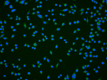
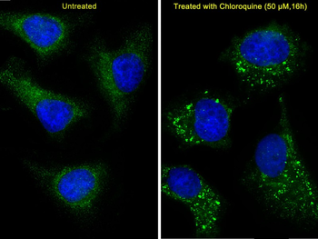
IF
Reactivity
Human
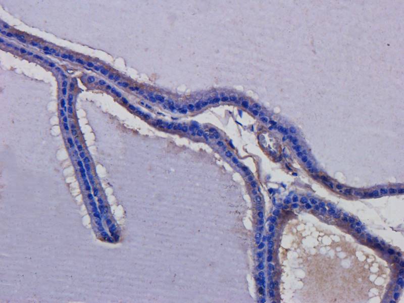
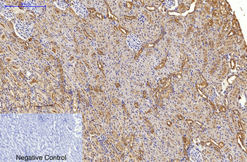
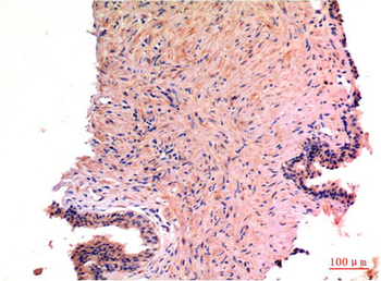
IHC-P image of rat prostate tissue using LC3 antibody (dilution of primary antibody at 2.5 ug/ml)
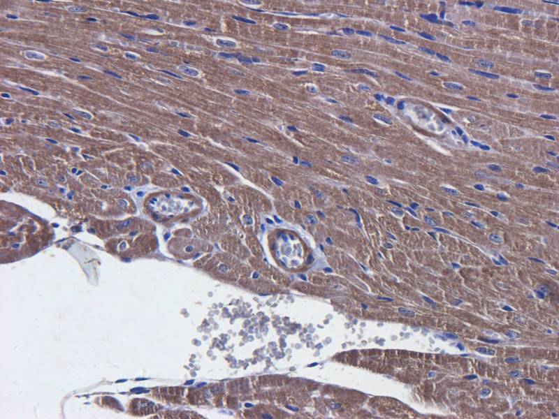
Immunohistochemical staining of mouse heart tissue using LC3 antibody (dilution of primary antibody - 2.5 ug/ml)
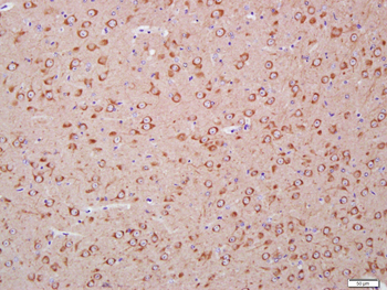
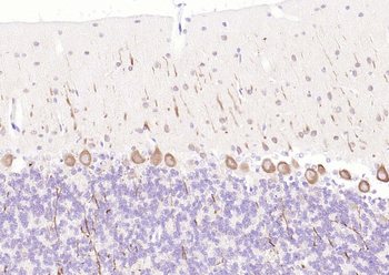
Immunohistochemical staining of rat spinal cord tissue using anti- LC3 (dilution of primary antibody - 2.5 ug/ml)
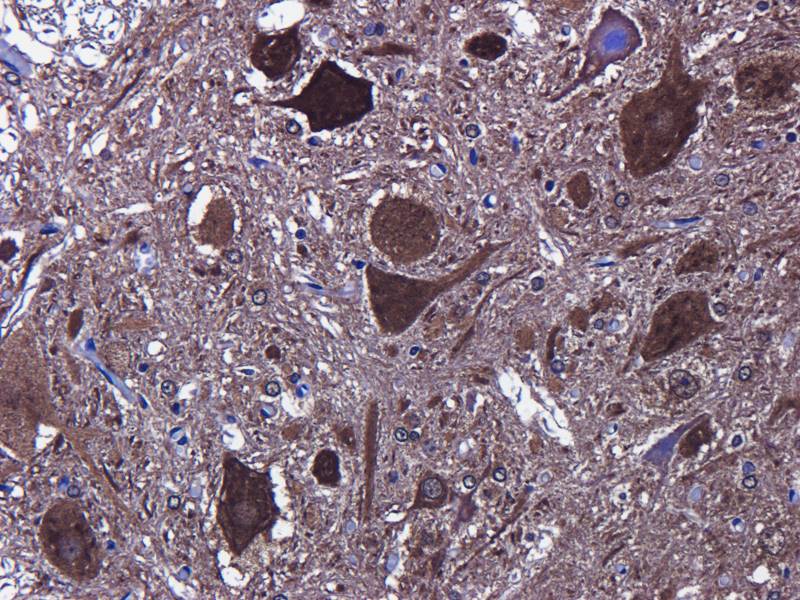
IHC-P staining of rat spinal cord tissue using LC3 antibody (dilution at 2.5 ug/ml)
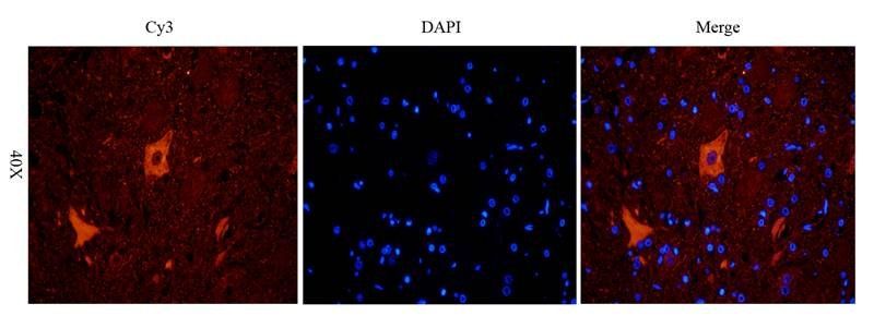
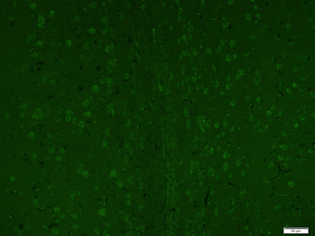
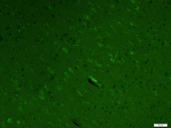
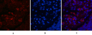
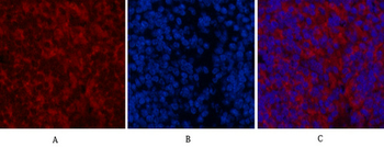
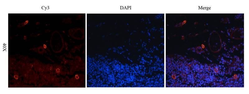
Immunofluorescence image of rat spinal cord tissue using anti- LC3 (dilution at 2.5 ug/ml)
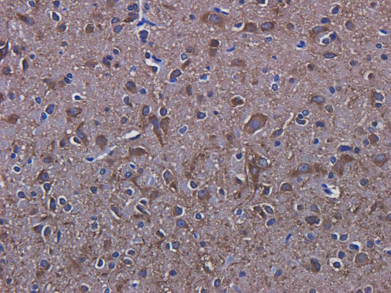
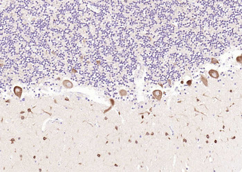
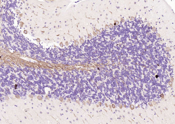
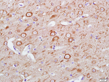
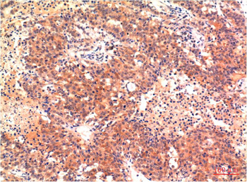
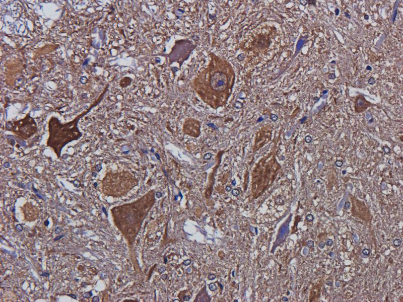
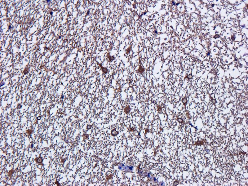
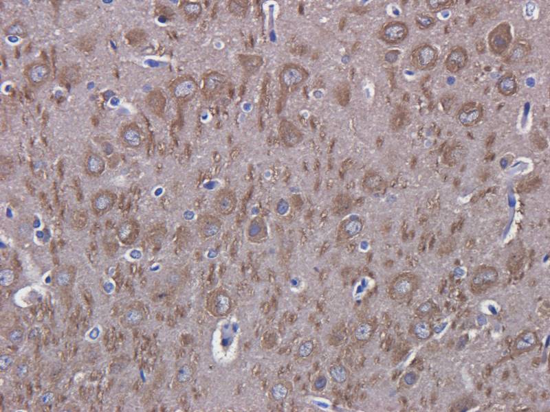
Immunohistochemical staining of paraffin embedded rat brain tissue using LC3 antibody (primary antibody at 2.5 ug/ml)
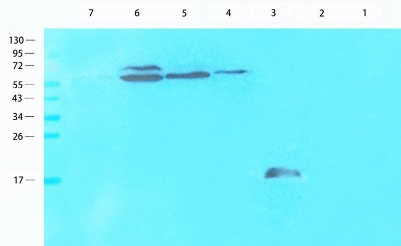
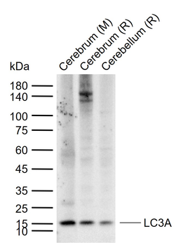
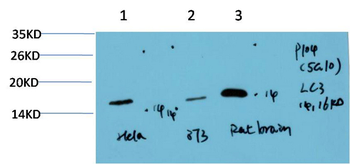
WB analysis of U251 cells (lane 1), 293T cells (lane 2), mouse brain (lane 3), rat spinal cord (lane 4), rat heart (lane 5), mouse uterus (lane 6), rat bladder (lane 7) using LC3 antibody (1 ug/ml)
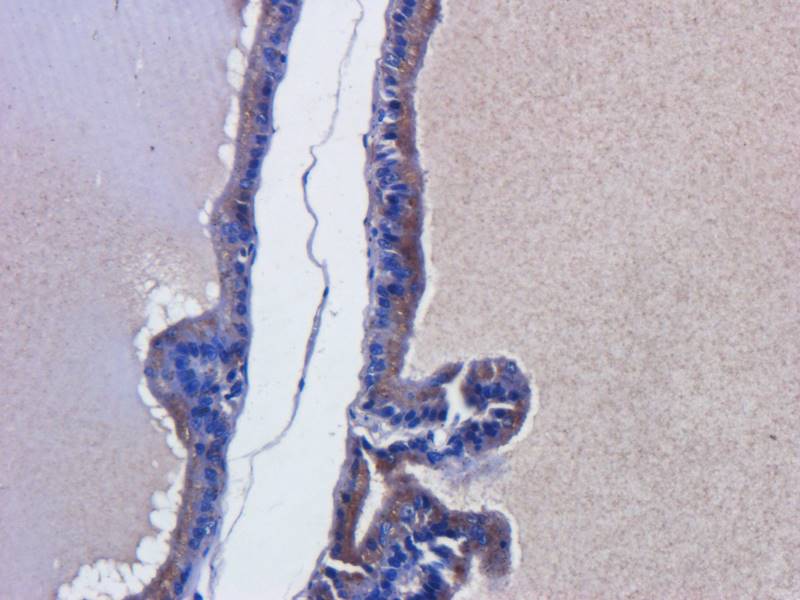
Immunohistochemical staining of paraffin embedded rat prostate tissue using LC3 antibody (primary antibody at 2.5 ug/ml)
IF image of rat thymus tissue using LC3 antibody (primary antibody at 2.5 ug/ml)
IHC-P image of rat spinal cord tissue using LC3 antibody (dilution of primary antibody at 2.5 ug/ml)
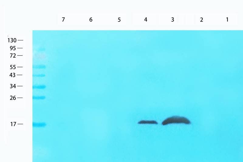
Wesstern blot analysis of U251 cells (lane 1), 293T cells (lane 2), mouse brain (lane 3), rat spinal cord (lane 4), rat heart (lane 5), mouse uterus (lane 6), rat bladder (lane 7) using LC3 antibody (1 ug/ml)
IHC-P staining of rat brain tissue using anti- LC3 (dilution at 2.5 ug/ml)
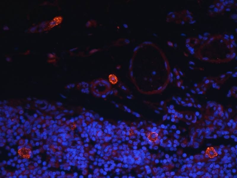
IF analysis of rat thymus tissue using LC3 antibody (dilution of primary antibody at 2.5 ug/ml)
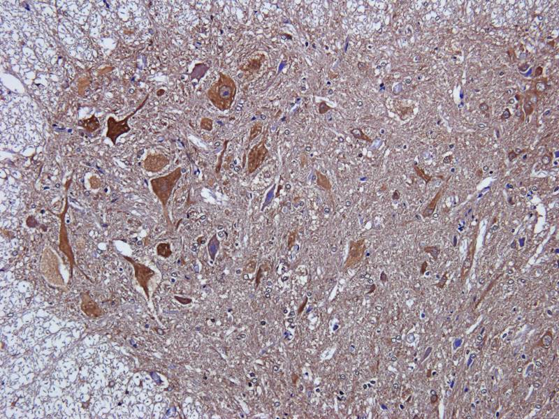
Immunohistochemical staining of rat spinal cord tissue using anti- LC3 (dilution of primary antibody - 2.5 ug/ml)
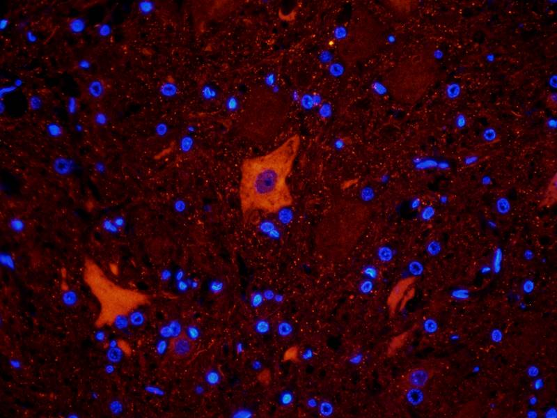
Immunofluorescence analysis of rat spinal cord tissue using LC3 antibody (dilution of primary antibody - 2.5 ug/ml)
- Item 1 of 10
LC3A Mouse Monoclonal Antibody [orb499565]
ICC, IF, IHC-Fr, IHC-P, WB
Mouse, Rat
Human, Mouse, Rat
Mouse
Monoclonal
Unconjugated
200 μl, 100 μl, 50 μl - Item 1 of 12
LC3B Antibody, KO Validated [orb1274430]
IF, IHC, WB
Human, Mouse, Porcine, Rat
Rabbit
Polyclonal
Unconjugated
100 μl - Item 1 of 7
LC3A mouse Monoclonal Antibody(5G10) [orb1415568]
IF, IHC-P, WB
Human, Mouse, Rat
Mouse
Monoclonal
Unconjugated
100 μl - Item 1 of 7
Cleaved LC3A Antibody [orb1933527]
ICC, IF, IHC-P, WB
Rat, Zebrafish
Human, Mouse
Rabbit
Polyclonal
Unconjugated
400 μl - Item 1 of 7
Cleaved LC3A Antibody [orb33336]
ICC, IF, IHC-P, WB
Rat, Zebrafish
Human, Mouse
Rabbit
Polyclonal
Unconjugated
80 μl