You have no items in your shopping cart.
Cart summary
Item 1 of 4
Item 1 of 4
KRT8 Antibody / Cytokeratin 8
Catalog Number: orb749629

| Catalog Number | orb749629 |
|---|---|
| Category | Antibodies |
| Description | Cytokeratin 8 (CK8) belongs to the type II (or B or basic) subfamily of high molecular weight cytokeratins and exists in combination with cytokeratin 18 (CK18). CK8 is primarily found in the non-squamous epithelia and is present in majority of adenocarcinomas and ductal carcinomas. It is absent in squamous cell carcinomas. Hepatocellular carcinomas are defined by the use of antibodies that recognize only cytokeratin 8 and 18. CK8 exists on several types of normal and neoplastic epithelia, including many ductal and glandular epithelia such as colon, stomach, small intestine, trachea, and esophagus as well as in transitional epithelium. Anti-CK8 does not react with skeletal muscle or nerve cells. Epithelioid sarcoma, chordoma, and adamantinoma show strong positivity corresponding to that of simple epithelia (with antibodies against CK8, CK18 and CK19). Reportedly, anti-CK8 is useful for the differentiation of lobular (ring-like, perinuclear) from ductal (peripheral-predominant) carcinoma of the breast. |
| Species/Host | Mouse |
| Clonality | Monoclonal |
| Clone Number | B22.1 |
| Tested applications | FACS, IF, IHC-P, WB |
| Reactivity | Human |
| Isotype | Mouse IgG1, kappa |
| Immunogen | A cytoskeletal preparation from HeLa cells was used as the immunogen for the Cytokeratin 8 antibody. |
| Dilution range | Flow cytometry: 0.5-1ug/million cells,Immunofluorescence: 1-2ug/ml,Immunohistochemistry (FFPE): 0.5-1ug/ml for 30 min at RT,Western blot: 1-2ug/ml |
| Purity | Protein G affinity chromatography |
| Conjugation | Unconjugated |
| Formula | 0.2 mg/ml in 1X PBS with 0.1 mg/ml BSA (US sourced) and 0.05% sodium azide |
| Hazard Information | This Cytokeratin 8 antibody is available for research use only. |
| UniProt ID | P05787 |
| Storage | Store the Cytokeratin 8 antibody at 2-8°C (with azide) or aliquot and store at -20°C or colder (without azide). |
| Buffer/Preservatives | 0.2 mg/ml in 1X PBS with 0.1 mg/ml rAlbumin (US sourced) and 0.05% sodium azide |
| Note | For research use only |
| Application notes | Optimal dilution of the Cytokeratin 8 antibody should be determined by the researcher.1. Staining of formalin-fixed tissues requires boiling tissue sections in 10mM Citrate buffer, pH 6.0, for 10-20 min followed by cooling at RT for 20 min2. The prediluted format is supplied in a dropper bottle and is optimized for use in IHC. After epitope retrieval step (if required), drip mAb solution onto the tissue section and incubate at RT for 30 min. |
| Expiration Date | 12 months from date of receipt. |
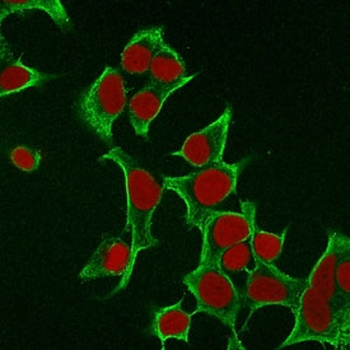
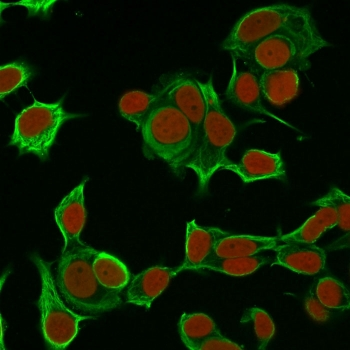
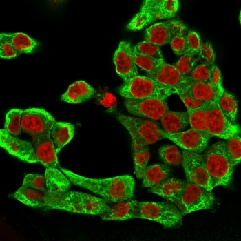
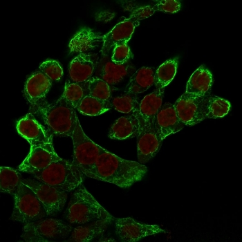
Immunofluorescent staining of permeabilized human MCF7 cells with Cytokeratin 8 antibody (clone B22.1, green) and Reddot nuclear stain (red).
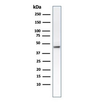
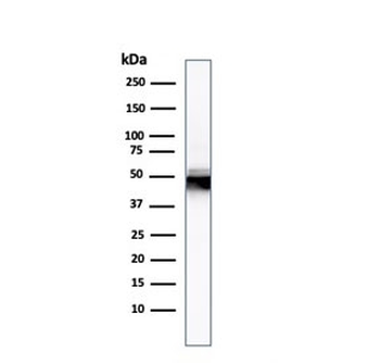
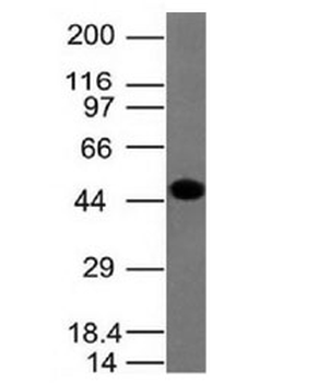
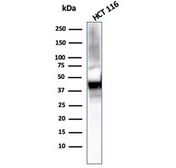
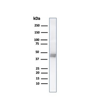
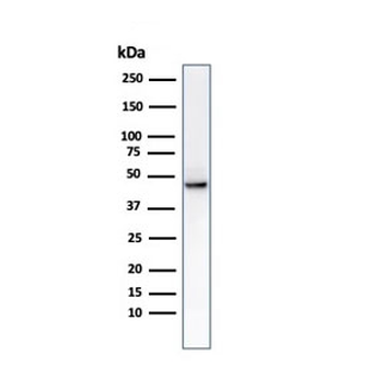
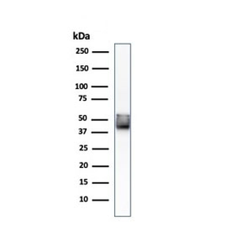
Western blot testing of human HCT-116 cell lysate with Cytokeratin 8 antibody (clone B22.1).
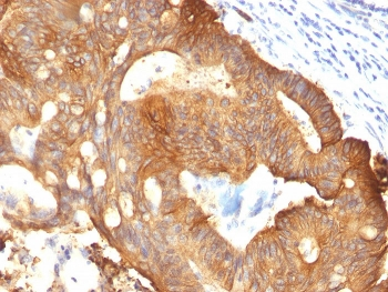
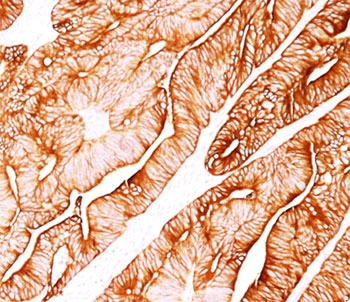
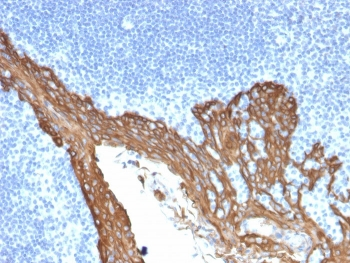
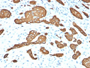
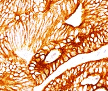
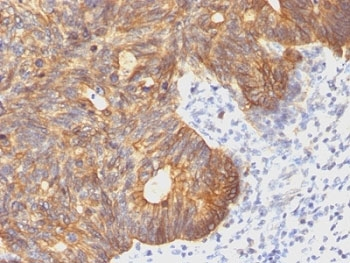
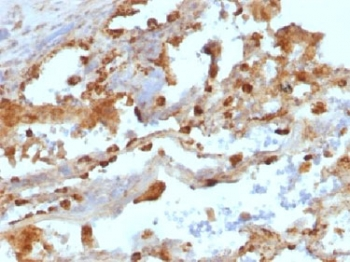
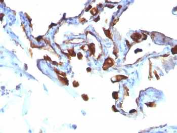
IHC: Formalin-fixed, paraffin-embedded colon carcinoma stained with Cytokeratin 8 antibody (clone B22.1). HIER: boil tissue sections in pH 9 10mM Tris with 1mM EDTA for 20 min and allow to cool before testing.
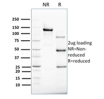
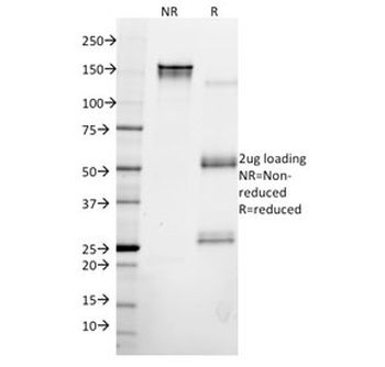
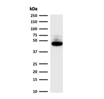
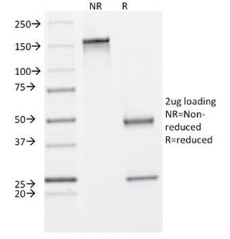
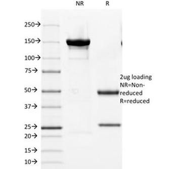
SDS-PAGE analysis of purified, BSA-free Cytokeratin 8 antibody (clone B22.1) as confirmation of integrity and purity.
- Item 1 of 6
KRT8 Antibody / Cytokeratin 8 [orb248434]
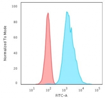
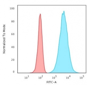
FACS, IF, IHC-P, WB
Human, Rat
Mouse
Monoclonal
Unconjugated
100 μg, 20 μg - Item 1 of 5
- Item 1 of 4
KRT8 Antibody / Cytokeratin 8 [orb749367]
FACS, IF, IHC-P, WB
Human
Mouse
Monoclonal
Unconjugated
100 μg, 20 μg - Item 1 of 4
KRT8 Antibody / Cytokeratin 8 [orb749630]
IF, IHC-P, WB
Human
Mouse
Monoclonal
Unconjugated
100 μg, 20 μg - Item 1 of 4
Submit a review
Filter by Rating

- 5 stars

- 4 stars
- 3 stars
- 2 stars
- 1 stars
