You have no items in your shopping cart.
Cart summary
Item 1 of 7
Item 1 of 7
JAM A pY280 antibody
Catalog Number: orb420323

| Catalog Number | orb420323 |
|---|---|
| Category | Antibodies |
| Description | JAM A pY280 antibody |
| Species/Host | Rabbit |
| Clonality | Polyclonal |
| Tested applications | ELISA, IF, WB |
| Reactivity | Human |
| Isotype | IgG |
| Immunogen | Affinity purified Anti-JAM A pY280 antibody was prepared from whole rabbit serum produced by repeated immunizations with a synthetic peptide corresponding to the c-term and phosphorylated at the tyrosine 280 position of Human JAM A protein. |
| Concentration | 1.0 mg/ml |
| Dilution range | ELISA: 5 µg/ml, IF: User Optimized, WB: 1 ug/ml |
| Form/Appearance | Liquid (sterile filtered) |
| Purity | Anti-JAM A pY280 is directed against human JAM A phosphorylated at the tyrosine 280 position. This product is an affinity purified antibody produced by immunoaffinity chromatography using phospho peptide coupled to agarose beads followed by solid phase adsorption(s) against non-phospho peptide to remove any unwanted reactivities. A BLAST analysis was used to suggest reactivity with this protein from human and feline based on 100% homology for the immunogen sequence. |
| Conjugation | Unconjugated |
| UniProt ID | 50848 |
| NCBI | NP_058642.1 |
| Storage | Store vial at -20° C or below prior to opening. This vial contains a relatively low volume of reagent (25 µL). To minimize loss of volume dilute 1:10 by adding 225 µL of the buffer stated above directly to the vial. Recap, mix thoroughly and briefly centrifuge to collect the volume at the bottom of the vial. Use this intermediate dilution when calculating final dilutions as recommended below. Store the vial at -20°C or below after dilution. Avoid cycles of freezing and thawing. |
| Buffer/Preservatives | 0.01% (w/v) Sodium Azide |
| Alternative names | rabbit anti-JAM A pY280 antibody, JAM-A, Junctiona Read more... |
| Note | For research use only |
| Application notes | This affinity purified antibody has been tested for use in ELISA, IF, and western blot. Specific conditions for reactivity should be optimized by the end user. Expect a band ~ 32.5 kDa in size corresponding to JAM A by western blotting in the appropriate cell lysate or extract. |
| Expiration Date | 12 months from date of receipt. |

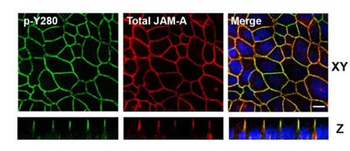
Confocal Immunofluorescence Microscopy of Rabbit Anti-JAM-A pY280 antibody in polarized epithelial cells. Tissue: T84 cells were grown on Transwell filters until confluent. Treatment: pervanadate. Fixation: 4% PFA. Permeabilization: 1% SDS. Costained Green: Anti-Phospho JAM-A Y280 Antibody, FITC conjugated secondary; Red: Anti-Total JAM-A, Alexa-conjugated secondary antibodies. Localization: tight junctions, seen in Confocal Z-stacks. Scale bar: 10 μm.
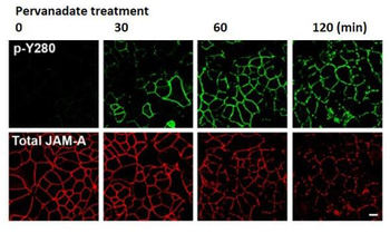
Confocal Immunofluorescence Microscopy of Rabbit Anti-JAM-A pY280 antibody of confluent (intestinal epithelial cells) IECs. Tissue: SK CO-15 cells. Treatment: pervanadate at time points 0, 30, 60, 120 mins. Fixation: 4% PFA. Permeabilization: 1% SDS. Costained Green: Anti-Phospho JAM-A Y280 Antibody, FITC conjugated secondary; Red: Anti-Total JAM-A, Alexa-conjugated secondary antibodies. Results: pervanadate treatment led to a time-dependent increase in phosphorylation of JAM-A Y280 that correlated with decreased localization of JAM-A at cell–cell contacts. Scale bar: 10 μm.

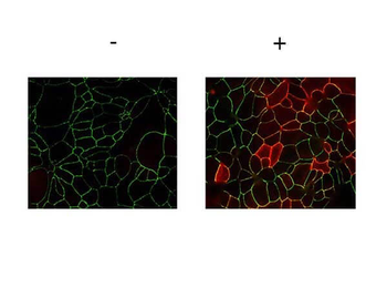
Immunofluorescence Microscopy of Rabbit anti-JAMA pY280 antibody. Tissue: T84 cells (untreated/treated). Fixation: 0.5% PFA. Antigen retrieval: not required. Primary antibody: JAMA pY280 antibody at 2 µg/mL for 1 hr at RT. Secondary antibody: Fluorescein rabbit secondary antibody at 1:10000 for 45 min at RT. Localization: JAMA pY280 is along the cell membrane and cell junction. Staining: JAMA pY280 as red fluorescent signal.

Immunofluorescence Microscopy of Rabbit Anti-JAM-A pY280 antibody. Tissue: T84 cells. Pretreatment: PP2. Treatment: Pervanadate. Fixation: 4% PFA. Permeabilization: 1% SDS. Costained Green: Anti-Phospho JAM-A Y280 Antibody, FITC conjugated secondary; Red: Anti-Total JAM-A, Alexa-conjugated secondary antibodies. Results: The Src family kinase inhibitor PP2 inhibits pervanadate-dependent phosphorylation of JAM-A Y280, as they reported to modulate tyrosine phosphorylation of junctional proteins and influence epithelial barrier function.

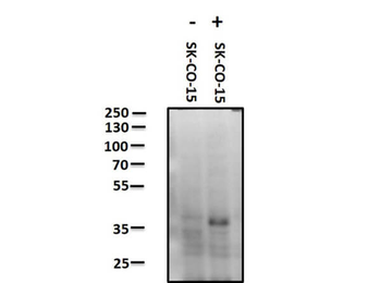
Western Blot of Rabbit anti-JAM A pY280 antibody. Lane 1: SK-CO-15 negative control. Lane 2: SK-CO-15 pervanadate treated positive control. Load: 10 µg per lane. Primary antibody: JAM A pY280 antibody at 1 ug/ml for overnight at 4°C. Secondary antibody: Peroxidase rabbit secondary antibody at 1:40000 for 30 min at RT. Block: orb348637 for 30 minutes at RT. Predicted/Observed size: ~ 32.5 kDa. JCYIA.

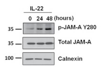
Western Blot of Rabbit Anti-JAM-A pY280 antibody with IL-22. Lysates: T84 cells. Treatment: hu rec. IL-22 at time points 0, 24, 48 hrs. Primary antibodies: JAM-A pY280, total JAM-A, or Calnexin. Calnexin was used as a loading control. Secondary antibody: horseradish peroxidase secondary antibody. Results: Exposure of IECs to other cytokines (IL-17A, IL-22, TNFα, or IFNγ) results in tyrosine phosphorylation of JAM-A at Y280 and a leaky barrier.

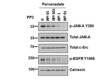
Western Blot of Rabbit Anti-JAM-A pY280 antibody with PP2. Lysates: T84 cells. Treatments: Pervanadate; PP2 at 0, 10nM, 100nM, 1 µM, 10 µM. Primary antibodies: p-JAM-A Y280, total JAM-A, total c-Src, p-EGFR Y1068, or Calnexin. p-EGFR Y1068 was used as a positive control for PP2. Calnexin was used as a loading control. Secondary antibody: horseradish peroxidase secondary antibody. Results: PP2 dose-dependent decrease in tyrosine phosphorylation of JAM-A Y280 following pervanadate treatment.
- Item 1 of 7


Submit a review
Filter by Rating

- 5 stars

- 4 stars
- 3 stars
- 2 stars
- 1 stars
