You have no items in your shopping cart.
Cart summary
Item 1 of 5
Item 1 of 5
IL6 antibody
Catalog Number: orb345214

| Catalog Number | orb345214 |
|---|---|
| Category | Antibodies |
| Description | IL6 antibody |
| Species/Host | Rabbit |
| Clonality | Polyclonal |
| Tested applications | ELISA, FC, IHC, WB |
| Reactivity | Mouse |
| Isotype | IgG |
| Immunogen | Anti-IL-6 is an IgG fraction antibody prepared from rabbit antiserum after repeated immunizations with recombinant mouse IL-6 protein produced in E.coli. |
| Concentration | 1.0 mg/mL |
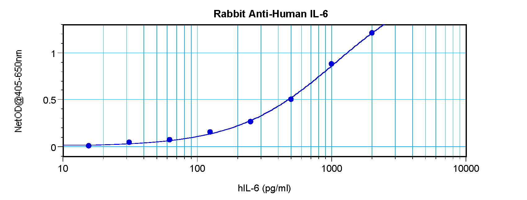
| Dilution range | ELISA: 1:10,000, IHC: User Optimized, WB: 1:1000 |
| Form/Appearance | Lyophilized |
| Purity | This product is an IgG fraction antibody purified from monospecific antiserum by a multi-step process which includes delipidation, salt fractionation and ion exchange chromatography followed by extensive dialysis against the buffer stated above. This antibody is specific for mouse IL-6 protein. A BLAST analysis was used to suggest cross-reactivity with IL-6 from mouse sources based on 100% homology with the immunizing sequence. Cross-reactivity with IL-6 from other sources has not been determined. |
| Conjugation | Unconjugated |
| UniProt ID | P08505 |
| NCBI | NP_112445.1 |
| Storage | Store anti-IL-6 at 4° C prior to restoration. For extended storage aliquot contents and freeze at -20° C or below. Avoid cycles of freezing and thawing. Centrifuge product if not completely clear after standing at room temperature. This product is stable for several weeks at 4° C as an undiluted liquid. Dilute only prior to immediate use. |
| Buffer/Preservatives | None |
| Alternative names | rabbit anti-IL-6 antibody, rabbit anti-Interleukin Read more... |
| Note | For research use only |
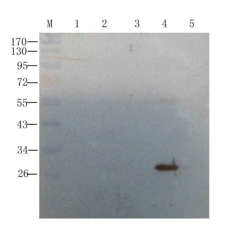
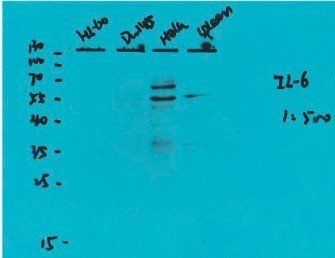
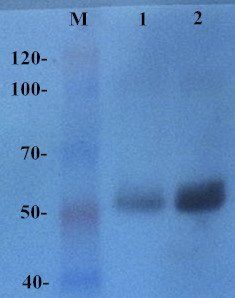
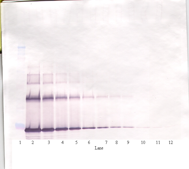
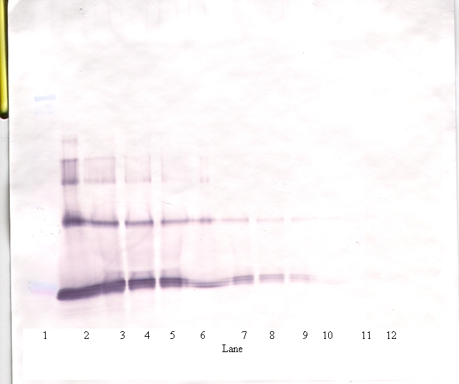
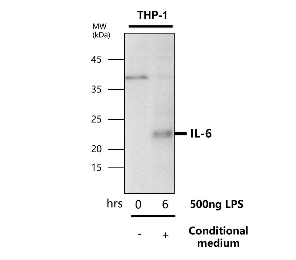
| Application notes | This purified antibody has been tested for use in Immunohistochemistry and western blotting and is suitable for ELISA. Specific conditions for reactivity should be optimized by the end user. Expect a band approximately 21.7 kDa in size corresponding to the mature 187 amino acid mouse IL-6 protein by western blotting in appropriate cell lysate or extract. |
| Expiration Date | 12 months from date of receipt. |
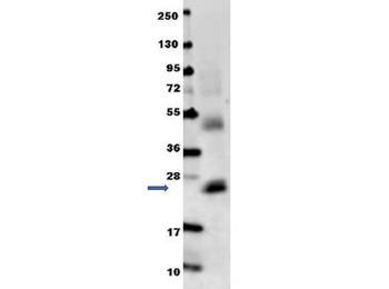
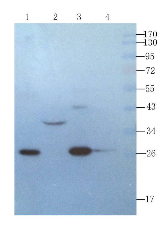
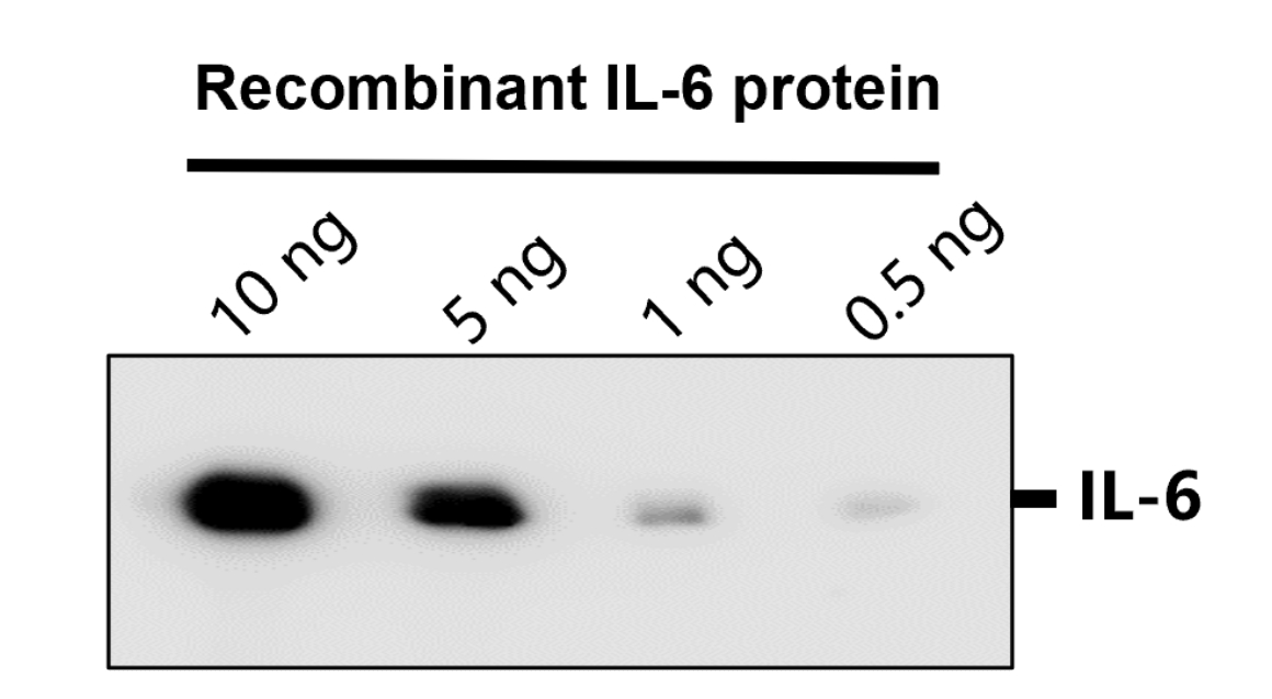
Anti-mouse IL-6 antibody in western blot shows detection of recombinant mouse IL-6 raised in E.coli. Recombinant truncated protein (0.1 µg, 21.7 kDa) was loaded on to an SDS-PAGE gel, and after separation, transferred to nitrocellulose. The membrane was blocked with 1% BSA in TBST for 30 min at RT, followed by incubation with Biorbyt's Anti-Mouse IL-6 antibody diluted 1:1000 in 1% BSA in TBST overnight at 4°C. After washes, the blot was reacted with secondary antibody Dylight™ 649 Conjugated Anti-Rabbit IgG (H&L) (Goat) Antibody diluted 1:20000 in blocking buffer (p/n orb348637) for 30 min at RT.
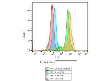
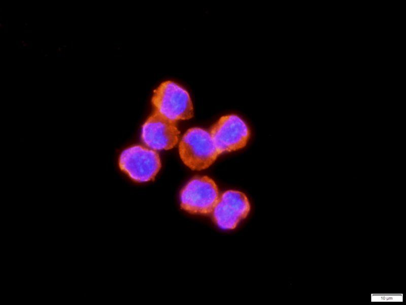
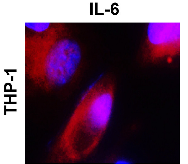
Flow Cytometry Results of Rabbit Anti-Mouse IL6 Antibody in human THP-1 cell line. The orange histogram represents the THP-1 cells that were activated with 100 ng/mL LPS for 24 hours. The green histogram are untreated THP-1 cells. These two populations were intracellularly stained for 30 minutes at 4°C in 1× BD Perm/Wash™ buffer. The primary stain was a 1:100 dilution of the Anti-Mouse IL-6 (RABBIT) Polyclonal Antibody (p/n orb345214) and the secondary stain was the Anti-RABBIT IgG (H&L) (GOAT) Antibody Fluorescein Conjugated (p/n orb347666 [1:400 dilution of 2 mg/mL]). The secondary stain was for 30 minutes at 4°C and was kept protected from light. The blue histogram is the THP-1 cells that were untreated and only stained with the secondary antibody. The red histogram is the untreated THP-1 cells that were not stained. Prior to staining, the cells for all conditions were permeabilized with BD Fixation/Permeabilization ™ solution for 20 minutes at 4°C. All washes and stains were performed in the BD 1× Perm/Wash™ buffer.
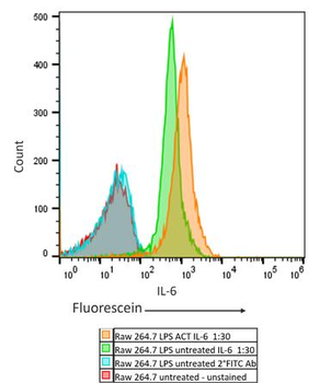
Flow Cytometry Results of Rabbit Anti-Mouse IL6 Antibody in mouse Raw 264.7 cell line. The orange histogram represents the Raw 264.7 murine cells that were activated with 100 ng/mL LPS for 24 hours. The green histogram are untreated Raw 264.7 cells. These two populations were intracellularly stained for 30 minutes at 4°C in 1× BD Perm/Wash™ buffer. The primary stain was a 1:30 dilution of the Anti-Mouse IL-6 (RABBIT) Polyclonal Antibody (p/n orb345214) and the secondary stain was the Anti-RABBIT IgG (H&L) (GOAT) Antibody Fluorescein Conjugated (p/n orb347666 [1:400 dilution of 2 mg/mL]). The secondary stain was for 30 minutes at 4°C and was kept protected from light. The blue histogram is the Raw 264.7 murine cells that were untreated and only stained with the secondary antibody. The red histogram is the untreated Raw 264.7 murine cells that were not stained. Prior to staining, the cells for all conditions were permeabilized with BD Fixation/Permeabilization ™ solution for 20 minutes at 4°C. All washes and stains were performed in the BD 1× Perm/Wash™ buffer.
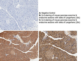
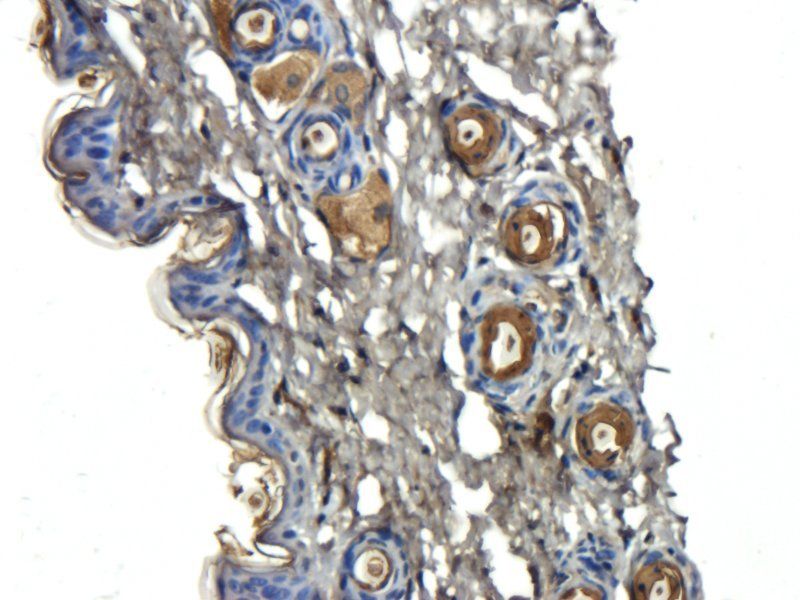
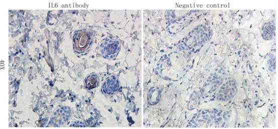
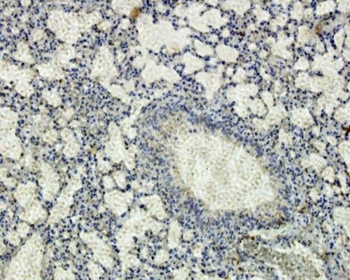
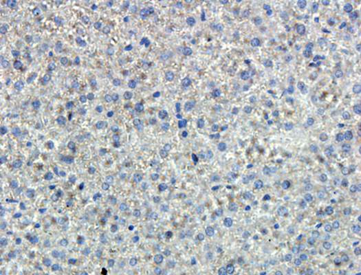
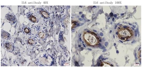
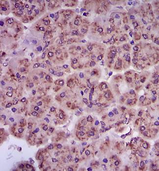
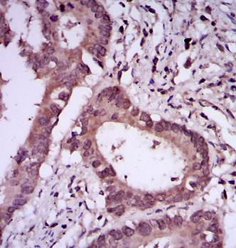
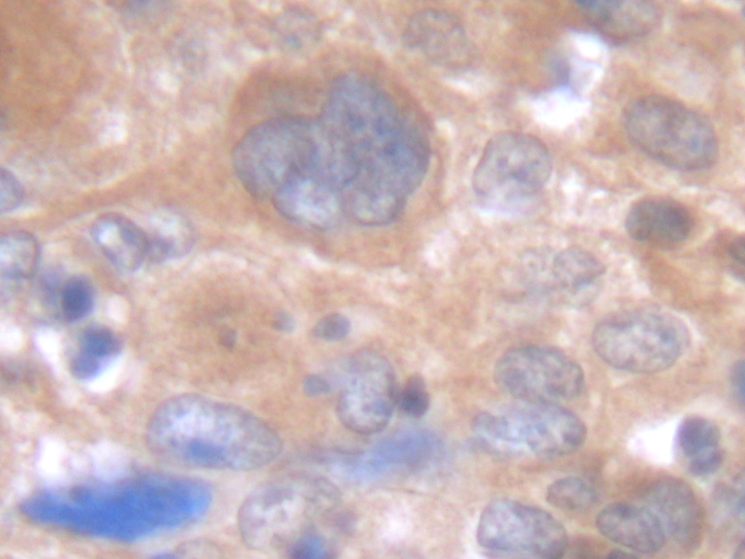
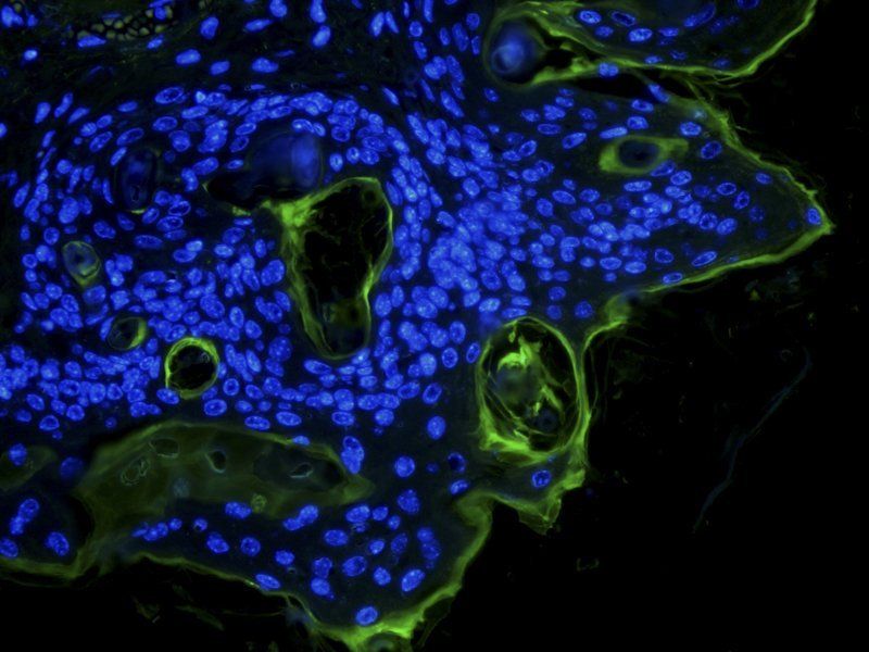
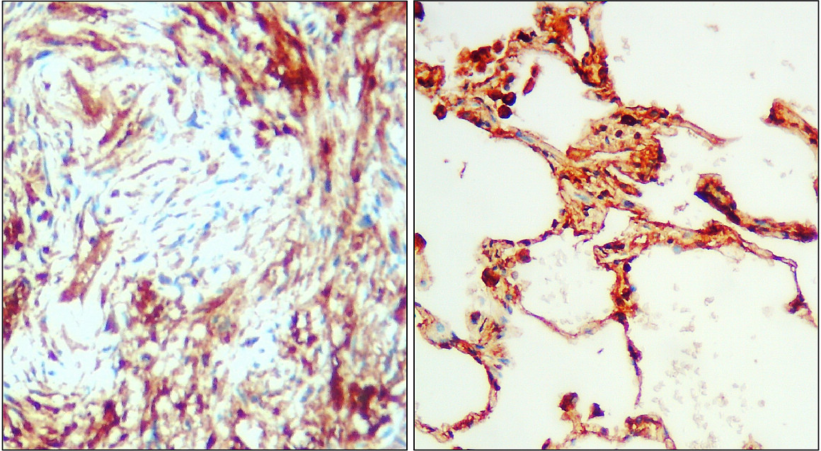
Immunohistochemistry with anti-IL-6 antibody showing cytoplasmic IL-6 staining in mouse pancreas exocrine and endocrine sections with islets of Langerhans at 10x and 20x (B & C). Formalin fixed/paraffin embedded tissue sections were subjected to antigen retrieval with E1 retrieval solution for 20 min and then incubated with rabbit anti-mouse IL-6 antibody at 1:50 dilution for 60 minutes. Biotinylated Anti-rabbit secondary antibody was used at 1:200 dilution to detect primary antibody. The reaction was developed using streptavidin-HRP conjugated compact polymer system and visualized with chromogen substrate, 3'3-diamino-benzidine substrate (DAB). The sections were then counterstained with hematoxylin to detect cell nuclei.
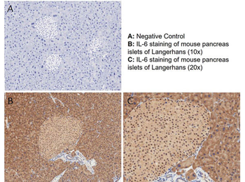
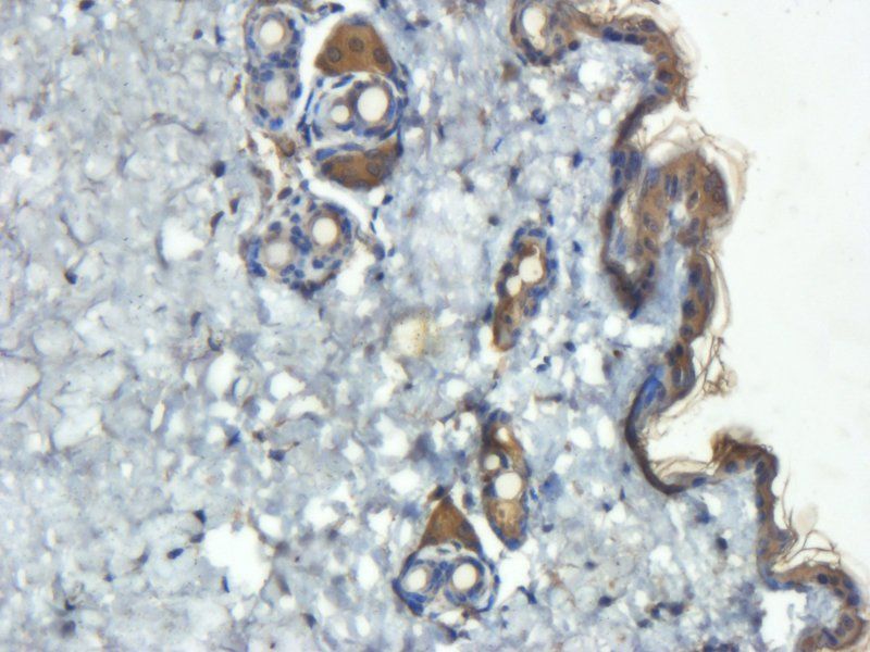
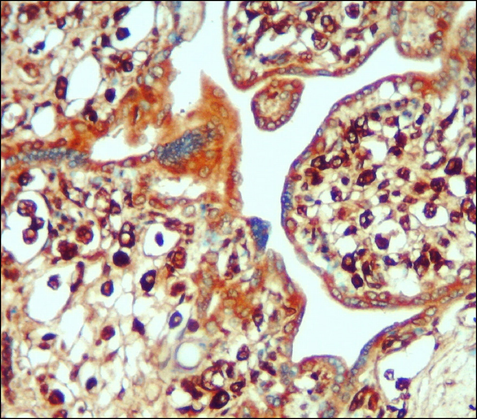
Immunohistochemistry with anti-IL-6 antibody showing positivity of islets of Langerhans (brown staining) and cytoplasmic staining in mouse pancreas at 10x and 20x (B & C). Formalin fixed/paraffin embedded tissue sections were subjected to antigen retrieval with E1 retrieval solution for 20 min and then incubated with rabbit anti-mouse IL-6 antibody at 1:50 dilution for 60 minutes. Biotinylated Anti-rabbit secondary antibody was used at 1:200 dilution to detect primary antibody. The reaction was developed using streptavidin-HRP conjugated compact polymer system and visualized with chromogen substrate, 3'3-diamino-benzidine substrate (DAB). The sections were then counterstained with hematoxylin to detect cell nuclei.
- Item 1 of 17
- Item 1 of 7
- Item 1 of 5
- Item 1 of 4
- Item 1 of 5

Submit a review
Filter by Rating

- 5 stars

- 4 stars
- 3 stars
- 2 stars
- 1 stars
