You have no items in your shopping cart.
Cart summary
Item 1 of 3
Item 1 of 3
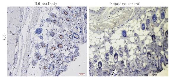
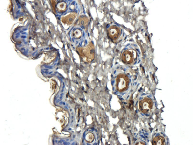
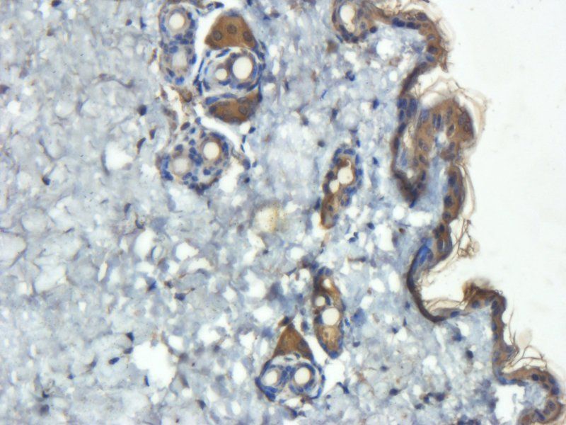
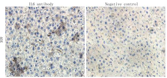
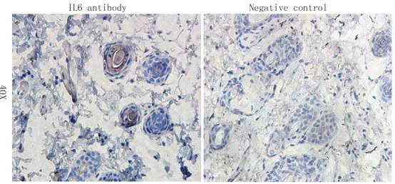
IL6 antibody
Catalog Number: orb345103

| Catalog Number | orb345103 |
|---|---|
| Category | Antibodies |
| Description | IL6 antibody |
| Species/Host | Mouse |
| Clonality | Monoclonal |
| Clone Number | 33A12.G9 |
| Tested applications | ELISA, FC, Multiplex Assay, WB |
| Reactivity | Human |
| Isotype | IgG1 |
| Immunogen | This Protein A purified monoclonal antibody was produced in mouse by repeated immunizations with mature full length recombinant human IL-6 produced in E.coli followed by hybridoma development. |
| Concentration | 1.0 mg/mL |
| Dilution range | ELISA: 1:10,000, FC: 0.5 mg/mL, WB: 1:1000 |
| Form/Appearance | Liquid (sterile filtered) |
| Purity | This purified antibody detects recombinant and native IL-6 present in body fluids and cell supernatants in various assays (ie. IL-1 stimulated IL-6 production from fibroblasts). In Western blot analysis of natural cell products or human body fluids, multiple bands of IL-6 will appear due to the variable amount of glycosylation on the molecule. |
| Conjugation | Unconjugated |
| UniProt ID | P05231 |
| NCBI | NP_000591.1 |
| Storage | Store vial at -20° C or below prior to opening. This vial contains a relatively low volume of reagent (25 µL). To minimize loss of volume dilute 1:10 by adding 225 µL of the buffer stated above directly to the vial. Recap, mix thoroughly and briefly centrifuge to collect the volume at the bottom of the vial. Use this intermediate dilution when calculating final dilutions as recommended below. Store the vial at -20°C or below after dilution. Avoid cycles of freezing and thawing. |
| Buffer/Preservatives | 0.01% (w/v) Sodium Azide |
| Alternative names | mouse Anti-IL-6 antibody, mouse anti-interleukin-6 Read more... |
| Note | For research use only |
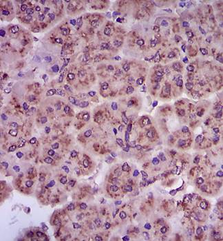
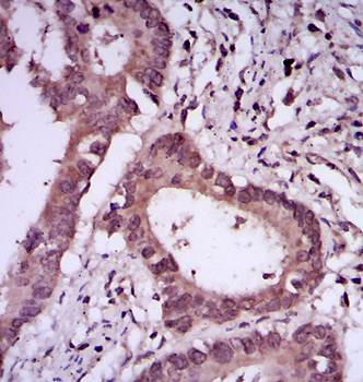
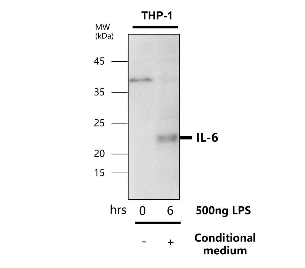
| Application notes | Anti-IL 6 antibody has been tested for use in ELISA, Flow Cytometry, and western blotting. Reactivity is also expected in neutralizations, radioimmunoassay and immunohistochemistry. The endotoxin content is estimated to be < 10 pg/µl by the LAL method. By western blot a band approximately 23.7 kDa in size corresponding to native human IL-6 protein is expected in the appropriate cell lysate or extract. Specific conditions for reactivity should be optimized by the end user. |
| Expiration Date | 12 months from date of receipt. |
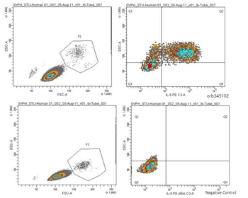
Anti-Human IL-6 Antibody - Flow Cytometry. Human PBMCs were stimulated with 1 ug/ml LPS and a transport inhibitor for 4-5 hours. Cells were then suspended in fixation buffer for 10-12 minutes and vortexed briefly. 1 mL of permeabilization buffer was added. 0.5mg of Anti-Human IL-6 Antibody was added (0.125 ug/ml control antibody) and incubated in the dark for 30 minutes. 1:100 of strep/PE was added and incubated for 30 minutes. LPS-stimulated samples were compared to unstimulated cells stained with strep/PE.
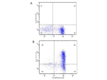
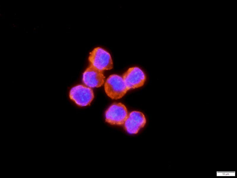
Flow Cytometry of Human anti-IL-6 antibody orb345102. Cells: human PBMC. Stimulation: Figure A: unstimulated; Figure B: 1 µg/mL LPS in a protein transport inhibitor for 5 hours. Staining: (surface) x-axis: anti-CD14, (intracellular) y-axis: anti-IL-6.
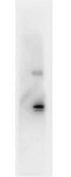
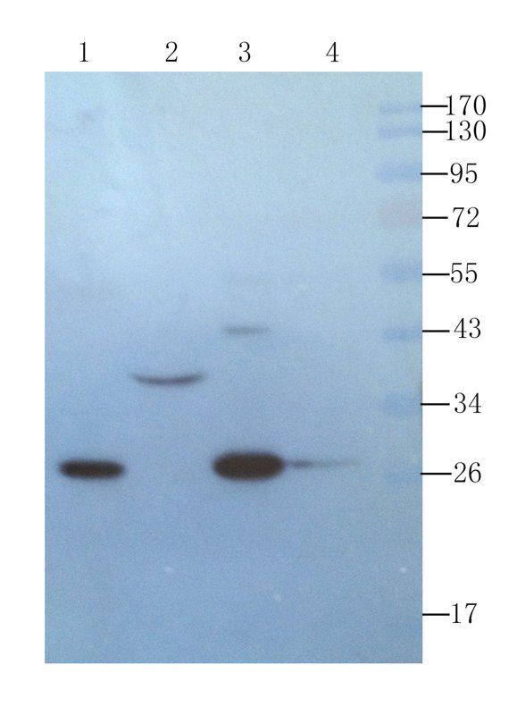
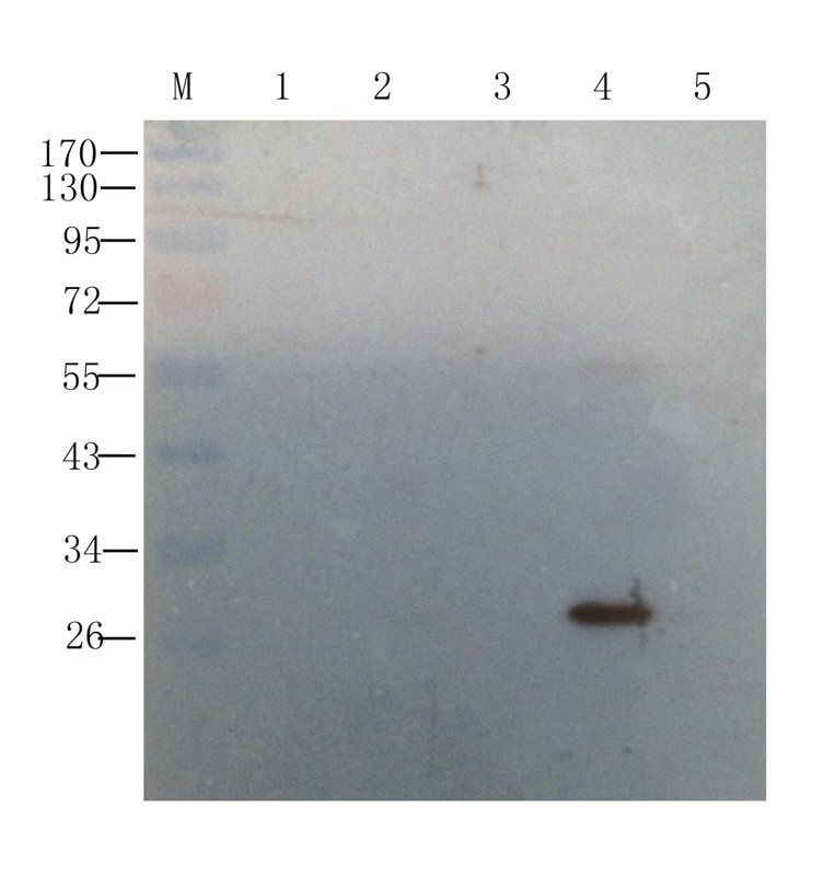
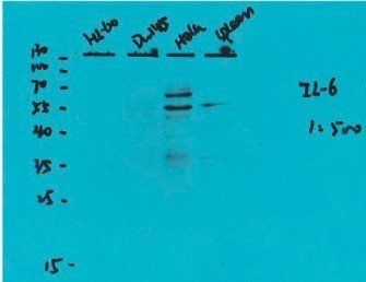
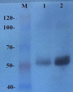
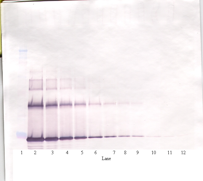
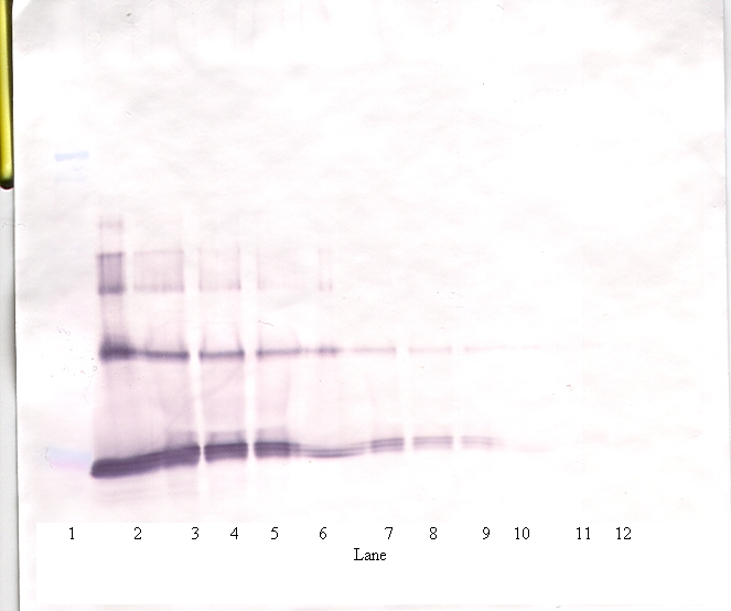
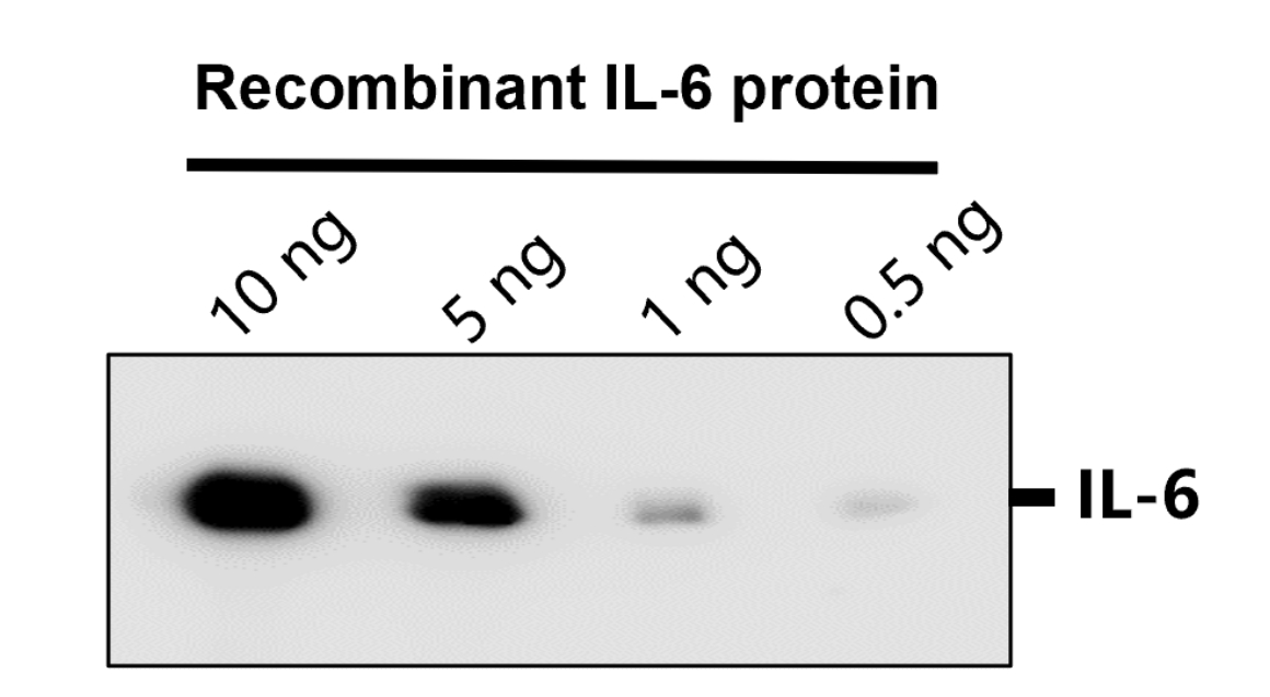
Western Blot showing detection of Human IL-6. 100 ng of Human IL-6 was run on a 4-20% gel and transferred to 0.45 µM nitrocellulose. After blocking with 1% BSA-TTBS (p/n orb348540, diluted to 1X) 30 min at 20°C, Anti-Human IL-6 (MOUSE) Antibody (p/n orb345102) was used at 1:1000 in 1% BSA-TTBS over night at 4°C. Peroxidase conjugated Rabbit Anti-mouse secondary antibody was used in Blocking Buffer for Fluorescent Western Blotting (p/n orb348637) at 1:40000 for 30 min at 20°C. Band indicates correct 17 kDa molecular weight position expected for Human IL-6.
- Item 1 of 17
- Item 1 of 7
- Item 1 of 5
- Item 1 of 4
- Item 1 of 5

Submit a review
Filter by Rating

- 5 stars

- 4 stars
- 3 stars
- 2 stars
- 1 stars
