You have no items in your shopping cart.
Cart summary
Item 1 of 3
Item 1 of 3
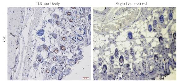
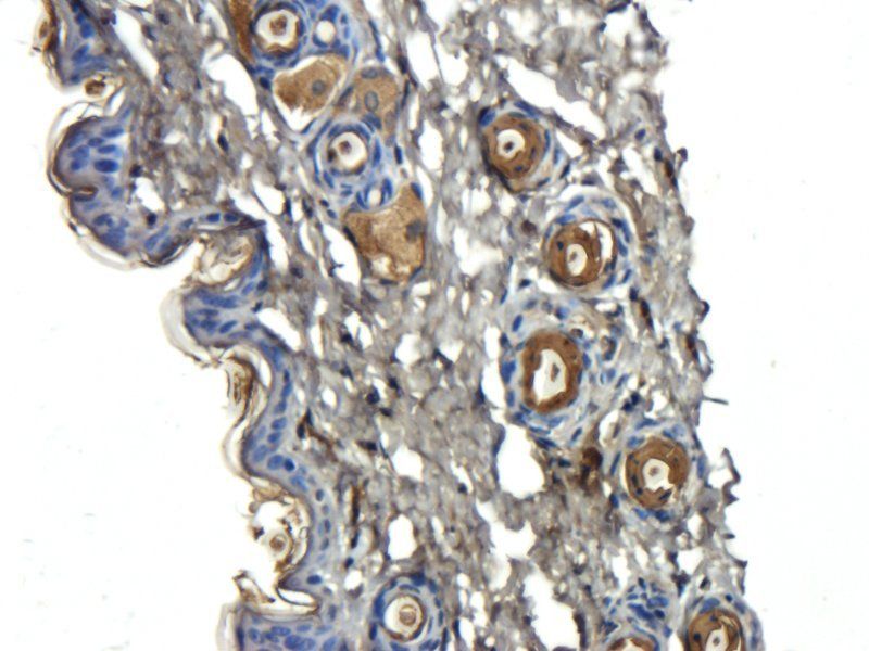
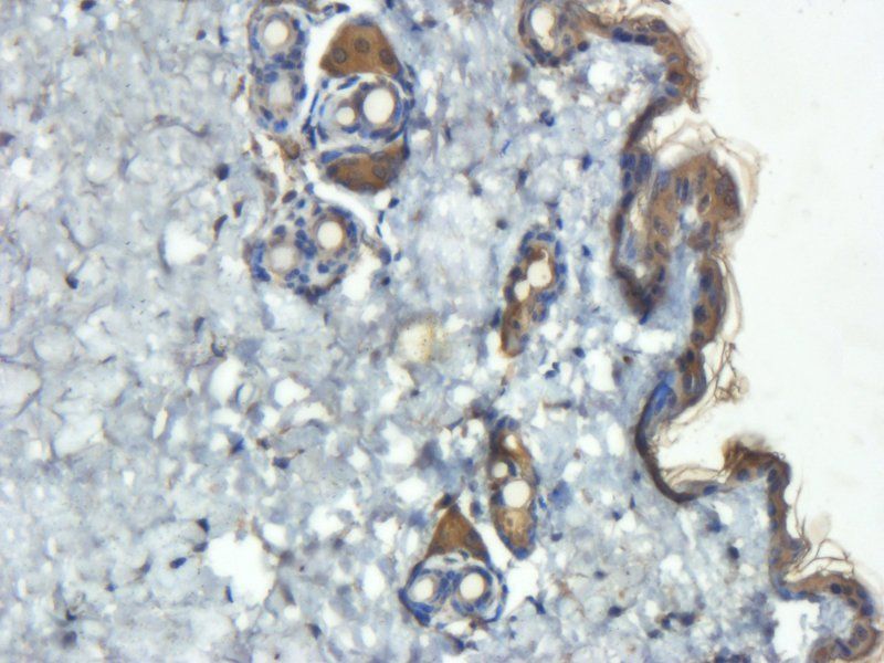
IL6 antibody
Catalog Number: orb345102

| Catalog Number | orb345102 |
|---|---|
| Category | Antibodies |
| Description | IL6 antibody |
| Species/Host | Mouse |
| Clonality | Monoclonal |
| Clone Number | 33A12.G9 |
| Tested applications | ELISA, FC, Multiplex Assay, WB |
| Reactivity | Human |
| Isotype | IgG1 |
| Immunogen | This Protein A purified IL-6 monoclonal antibody was produced in mouse by repeated immunizations with mature full length recombinant human IL-6 produced in E.coli followed by hybridoma development. |
| Concentration | 1.0 mg/mL |
| Dilution range | ELISA: 1:10,000, FC: 0.5 mg/mL, WB: 1:1000 |
| Form/Appearance | Liquid (sterile filtered) |
| Purity | This purified antibody detects recombinant and native IL-6 present in body fluids and cell supernatants in various assays (ie. IL-1 stimulated IL-6 production from fibroblasts). In Western blot analysis of natural cell products or human body fluids, multiple bands of IL-6 will appear due to the variable amount of glycosylation on the molecule. |
| Conjugation | Unconjugated |
| UniProt ID | P05231 |
| NCBI | NP_000591.1 |
| Storage | Store IL 6 antibody at -20° C . For extended storage aliquot contents and freeze at -20° C or below. Avoid cycles of freezing and thawing. Centrifuge product if not completely clear after standing at room temperature. This product is stable for several weeks at 4° C as an undiluted liquid. Dilute only prior to immediate use. |
| Buffer/Preservatives | 0.01% (w/v) Sodium Azide |
| Alternative names | mouse Anti-IL-6 antibody, mouse anti-interleukin-6 Read more... |
| Note | For research use only |
| Application notes | Anti-IL-6 antibody has been tested for use in ELISA, Flow Cytometry, and western blotting. Reactivity is also expected in neutralizations, radioimmunoassay and immunohistochemistry. The endotoxin content is estimated to be < 10 pg/µl by the LAL method. By western blot a band approximately 23.7 kDa in size corresponding to native human IL-6 protein is expected in the appropriate cell lysate or extract. Specific conditions for reactivity should be optimized by the end user. |
| Expiration Date | 12 months from date of receipt. |
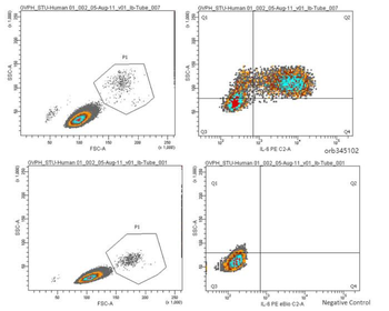
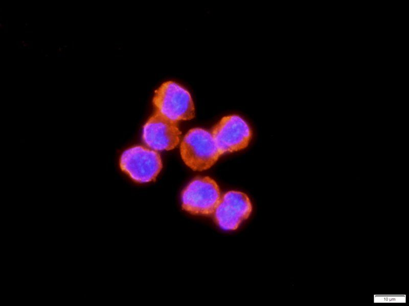
Anti-Human IL-6 Antibody - Flow Cytometry. Human PBMCs were stimulated with 1 ug/ml LPS and a transport inhibitor for 4-5 hours. Cells were then suspended in fixation buffer for 10-12 minutes and vortexed briefly. 1 mL of permeabilization buffer was added. 0.5mg of Anti-Human IL-6 Antibody was added (0.125 ug/ml control antibody) and incubated in the dark for 30 minutes. 1:100 of strep/PE was added and incubated for 30 minutes. LPS-stimulated samples were compared to unstimulated cells stained with strep/PE.
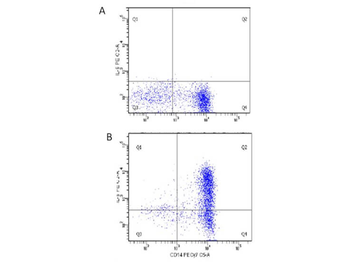
Flow Cytometry of Human anti-IL-6 antibody orb345102. Cells: human PBMC. Stimulation: Figure A: unstimulated; Figure B: 1 µg/mL LPS in a protein transport inhibitor for 5 hours. Staining: (surface) x-axis: anti-CD14, (intracellular) y-axis: anti-IL-6.
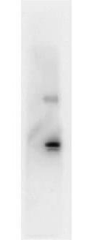
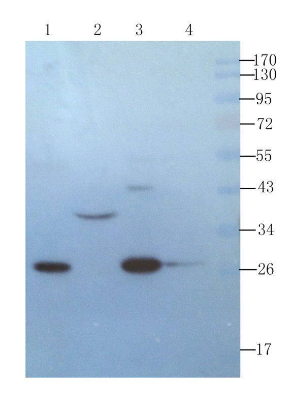
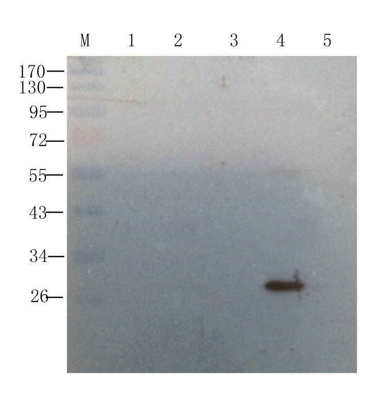
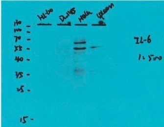
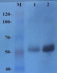
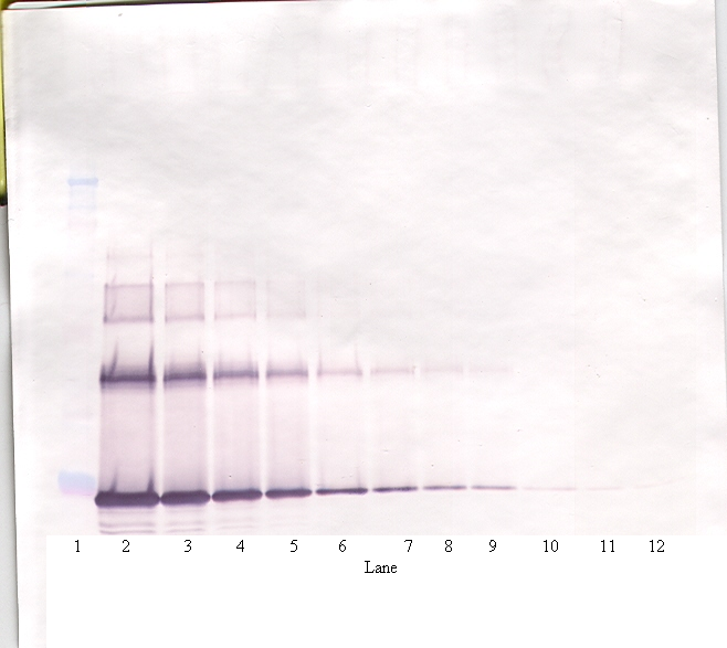
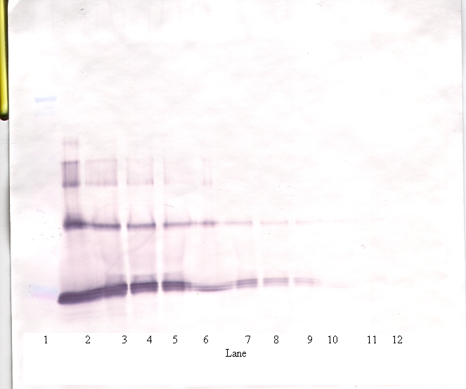
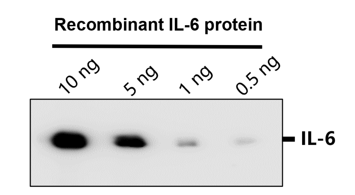
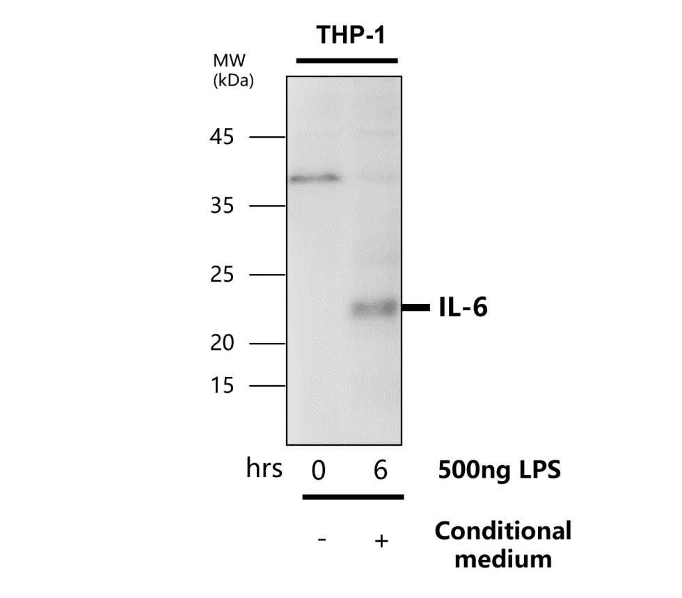
Western Blot showing detection of Human IL-6. 100 ng of Human IL-6 was run on a 4-20% gel and transferred to 0.45 µM nitrocellulose. After blocking with 1% BSA-TTBS (p/n orb348540, diluted to 1X) 30 min at 20°C, Anti-Human IL-6 (MOUSE) Antibody (p/n orb345102) was used at 1:1000 in 1% BSA-TTBS over night at 4°C. Peroxidase conjugated Rabbit Anti-mouse secondary antibody was used in Blocking Buffer for Fluorescent Western Blotting (p/n orb348637) at 1:40000 for 30 min at 20°C. Band indicates correct 17 kDa molecular weight position expected for Human IL-6.
- Item 1 of 17
- Item 1 of 7
- Item 1 of 5
- Item 1 of 4
- Item 1 of 5

Submit a review
Filter by Rating

- 5 stars

- 4 stars
- 3 stars
- 2 stars
- 1 stars
