You have no items in your shopping cart.
Cart summary
Item 1 of 1
IL-17A Antibody Biotin Conjugated
Catalog Number: orb345174

| Catalog Number | orb345174 |
|---|---|
| Category | Antibodies |
| Description | IL-17A antibody (Biotin) |
| Species/Host | Rabbit |
| Clonality | Polyclonal |
| Tested applications | ELISA, IHC, WB |
| Reactivity | Human |
| Isotype | IgG |
| Immunogen | This purified antibody was prepared from whole rabbit serum produced by repeated immunizations with full length recombinant human IL17-A protein. |
| Antibody Type | Primary Antibody |
| Concentration | 1.0 mg/ml |
| Dilution range | ELISA: 1:20,000-1:100,000, IHC: 1:1,000-1:5,000, WB: 1:2,000-1:10,000 |
| Form/Appearance | Lyophilized |
| Purity | This purified antibody has been heated to 56°C for 30 minutes. In ELISA and other immunoreactive assays, this antibody will recognize both native and recombinant human IL17-A in cell supernatants and certain body fluids. A control of similarly diluted normal rabbit IgG is recommended. |
| Conjugation | Biotin |
| UniProt ID | Q16552 |
| NCBI | AAH662531.1 |
| Storage | Store vial at 4° C prior to restoration. For extended storage aliquot contents and freeze at -20° C or below. Avoid cycles of freezing and thawing. Centrifuge product if not completely clear after standing at room temperature. This product is stable for several weeks at 4° C as an undiluted liquid. Dilute only prior to immediate use. |
| Buffer/Preservatives | 0.01% (w/v) Sodium Azide |
| Alternative names | rabbit anti-Interleukin-17A biotin conjugated anti Read more... |
| Note | For research use only |
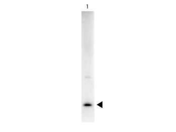
| Application notes | This purified antibody has been tested in western blotting. By western blot a band approximately 30.3 kDa in size corresponding to human IL17-A protein is expected in the appropriate cell lysate or extract. Specific conditions for reactivity should be optimized by the end user. |
| Expiration Date | 12 months from date of receipt. |
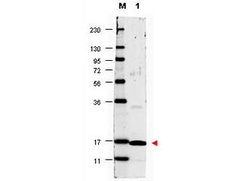
Western blot using Biorbyt's anti-Human IL17-A antibody shows detection of a band ~17 kDa in size corresponding to recombinant human IL17-A (lane 1). Molecular weight markers are also shown (M). After transfer, the membrane was blocked overnight with 3% BSA in TBS followed by reaction with primary antibody at a 1:1000 dilution. Detection occurred using DyLight 649 conjugated anti-Rabbit IgG secondary antibody diluted 1:20000 in blocking buffer (p/n orb348637).
- Item 1 of 1
- Item 1 of 1

- Item 1 of 1
- Item 1 of 1