You have no items in your shopping cart.
Cart summary
Item 1 of 3
Item 1 of 3
IL-1 Beta Antibody
Catalog Number: orb345217

| Catalog Number | orb345217 |
|---|---|
| Category | Antibodies |
| Description | IL1 beta antibody |
| Species/Host | Rabbit |
| Clonality | Polyclonal |
| Tested applications | ELISA, IF, IHC, IP, WB |
| Reactivity | Mouse |
| Isotype | IgG |
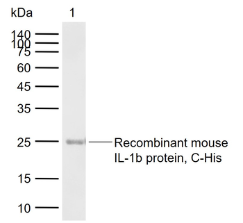
| Immunogen | This antibody was prepared by repeated immunizations with recombinant mouse IL-1ß produced in E.coli. The MW of recombinant mouse IL-1ß was 17 kDa. |
| Antibody Type | Primary Antibody |
| Concentration | 1.0 mg/mL |
| Dilution range | ELISA: 1:1,000 - 1:5,000, IHC: 1:50-1:250, IF: 1:50-1:250, IP: 1:200-1:800, WB: 1:500 - 1:2,000 |
| Form/Appearance | Lyophilized |
| Purity | This is an IgG preparation of whole rabbit serum purified by DEAE fractionation. This antibody is primarily directed against mature, 17,000 MW mouse IL-1ß and is useful in determining its presence in various assays. The antibody does not recognize human IL-1ß or mouse IL-1α based on a neutralization assay. In ELISA formats and other immunoreactive assays, reactivity occurs with rat IL-1ß. This antibody will recognize 10% of the non-denatured (native) precursor 31,000 MW mouse IL-1ß containing samples but will primarily detect all of the 17,000 MW mature molecule. However, in immunoblot analysis, the usual procedure of heating the sample in SDS with or without reducing agents will facilitate denaturing of the 31,000 MW IL- 1ß precursor molecule. Denatured 31,000 precursor IL-1ß will be recognized by this antibody. |
| Conjugation | Unconjugated |
| UniProt ID | P10749 |
| NCBI | CAA28637.1 |
| Storage | Store Anti-IL-1 beta antibody at 4° C prior to restoration. For extended storage aliquot contents and freeze at -20° C or below. Avoid cycles of freezing and thawing. Centrifuge product if not completely clear after standing at room temperature. This product is stable for several weeks at 4° C as an undiluted liquid. Dilute only prior to immediate use. |
| Buffer/Preservatives | None |
| Alternative names | rabbit anti-IL-1 beta antibody, rabbit anti-IL-1b Read more... |
| Note | For research use only |
| Application notes | Anti-Mouse IL-1ß has been tested for use in immunohistochemistry, immunoblotting and immunofluorescence. This antibody is useful in ELISA, neutralizations, radioimmunoassays, flow cytometry, and immunoprecipitation. It recognizes the 17,000 MW mature IL-1ß. For immunoblots, typically, IL-1ß is detected from supernatants or lysates of 2 x 10E6 endotoxin-stimulated peripheral blood mononuclear cells (PBMC). PBMC are stimulated for 24 hours with 1% (v/v) serum plus 10 ng/mL E.coli LPS. For immunoprecipitation pre-clearing the preparation with a non-specific Rabbit IgG (p/n 011-001-297) to reduce background is suggested. For immunohistochemistry either paraffin fixation or cryofixation can be used for sample preparation to stain intracellular IL-1ß. For ELISA use HRP Conjugated Anti-Rabbit IgG [H&L] (Goat) (611-1302) for detection. In ELISA formats this antibody is best used as the second antibody in combination with a monoclonal antibody as a capture antibody. This antibody is also useful for neutralization of mouse and rat IL-1ß activity in bioassays. It does not neutralize the biological activity IL-1α. It does not neutralize the biological activity of human or primate IL-1ß. For neutralization, it is recommended to incubate the sample with a dilution of the antibody for at least 4 hours before being tested. A control of similarly diluted normal rabbit IgG is recommended. This antibody can be used for FACS analysis. Caution should be exhibited as the F(c) domain of the rabbit IgG molecule may interact with cells non-specifically. |
| Expiration Date | 12 months from date of receipt. |
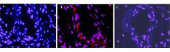
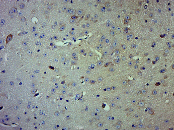
Immunofluorescence microscopy after staining of mouse carotid artery tissue with anti-Mouse IL-1ß antiserum diluted 1:50. Tissue sections were prepared after cryofixation. Reaction occurred at room temperature for 60' followed by washes and reaction with Rhodamine conjugated Gt-a-Rabbit IgG (p/n orb347644). Tissue was counterstained with bis-benzimide solution at 0.5 mg/ml in PBS for 15 min at room temperature. Panel A) shows no antibody staining of WT uninjured mouse carotid tissue. Panel B) shows anti-IL-1ß staining of cells after surgical injury of tissue. Panel C) shows no antibody staining of injured carotid tissue from an IL-1ß KO mouse.
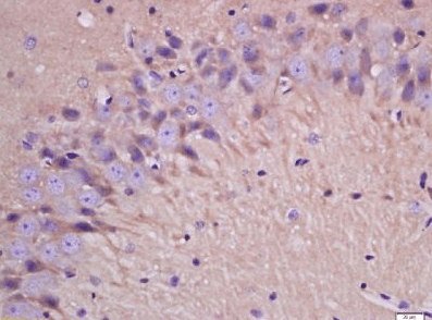
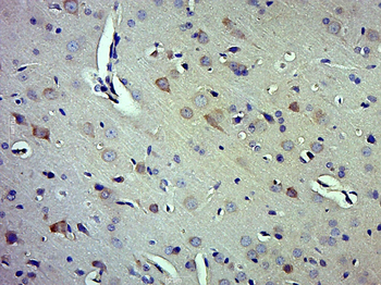
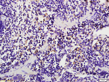
Immunohistochemistry of Rabbit anti-IL1Beta Antibody in Mouse Embryonic Kidney Tissue: Mouse Embryonic Kidney Fixation: FFPE buffered formalin 10% conc Ag Retrieval: Heat, Citrate pH6.2. Pressure Cooker Primary antibody: 2 ug/ml 1.5 hour @ room T Secondary Ab: MACH 1 HRP POLYMER 1:50 45" RT
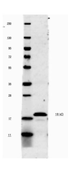
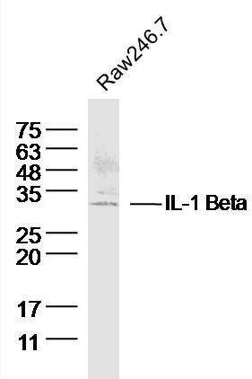
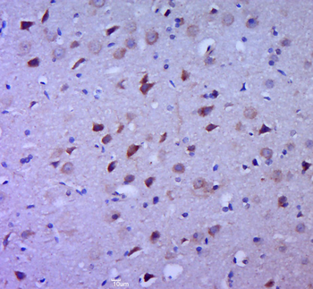
This antibody will recognize 10% of the non-denatured (native) precursor 31000 MW mouse IL-1ß containing samples but will primarily detect all of the 17000 MW mature molecule. However, in western blot analysis, the usual procedure of heating the sample in SDS with or without reducing agents will facilitate denaturing of the 31, 000 MW IL- 1ß precursor molecule. Denatured IL-1ß will have a 18 kDa band.
- Item 1 of 6
- Item 1 of 4
IL-1 Beta Rabbit Polyclonal Antibody [orb10903]
ELISA, FC, IF, IHC-Fr, IHC-P, WB
Mouse, Rat
Human, Mouse, Rat
Rabbit
Polyclonal
Unconjugated
100 μl, 50 μl, 200 μl - Item 1 of 5
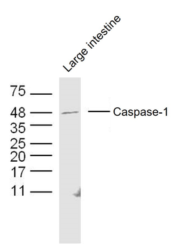
Caspase-1 P10 Rabbit Polyclonal Antibody [orb10232]
FC, IF, IHC-Fr, IHC-P
Rat
Human, Mouse, Rat
Rabbit
Polyclonal
Unconjugated
100 μl, 50 μl, 200 μl - Item 1 of 4
IL-1 Beta Rabbit Polyclonal Antibody [orb101745]
FC, IF, IHC-Fr, IHC-P, WB
Canine, Equine, Rabbit
Human, Mouse, Rat
Rabbit
Polyclonal
Unconjugated
50 μl, 100 μl, 200 μl - Item 1 of 5
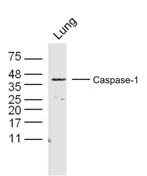
Caspase-1 p20 Rabbit Polyclonal Antibody [orb317579]
FC, IF, IHC-Fr, IHC-P, WB
Human, Rat
Human, Mouse, Rat
Rabbit
Polyclonal
Unconjugated
100 μl, 50 μl, 200 μl