You have no items in your shopping cart.
Cart summary
Item 1 of 6
Item 1 of 6
IDO antibody
Catalog Number: orb345211

| Catalog Number | orb345211 |
|---|---|
| Category | Antibodies |
| Description | IDO antibody |
| Species/Host | Mouse |
| Clonality | Monoclonal |
| Clone Number | 2E2.6 |
| Tested applications | ELISA, FC, IF, IHC, IP, Multiplex Assay, WB |
| Reactivity | Mouse |
| Isotype | IgG1 |
| Immunogen | IDO1 antibody was produced in mouse by repeated immunizations with mouse recombinant IDO1 protein followed by hybridoma development. |
| Concentration | 0.95 mg/mL |
| Dilution range | ELISA: 1:5000-1:50000, FC: 0.5-1x10^6 cells, IHC: User Optimized, IF: 1:50-1:100, IP: 10-100 µL, WB: 1:500-1:1500 |
| Form/Appearance | Liquid (sterile filtered) |
| Purity | Anti-IDO1 antibody was purified from concentrated tissue culture supernate by Protein G chromatography followed by extensive dialysis against the buffer stated above. IDO1 antibody is specific for mouse IDO1 protein. Mouse IDO1 does not react with human tissues. Cross-reactivity with IDO1 from other sources has not been determined. |
| Conjugation | Unconjugated |
| UniProt ID | P28776 |
| NCBI | NP_032350.1 |
| Storage | Store vial at -20° C or below prior to opening. This vial contains a relatively low volume of reagent (25 µL). To minimize loss of volume dilute 1:10 by adding 225 µL of the buffer stated above directly to the vial. Recap, mix thoroughly and briefly centrifuge to collect the volume at the bottom of the vial. Use this intermediate dilution when calculating final dilutions as recommended below. Store the vial at -20°C or below after dilution. Avoid cycles of freezing and thawing. |
| Buffer/Preservatives | 0.01% (w/v) Sodium Azide |
| Alternative names | mouse anti-IDO1 antibody, Ido, Indo, Indoleamine 2 Read more... |
| Note | For research use only |
| Application notes | Anti-IDO1 antibody has been tested for use in ELISA, Western Blot, IF, IHC, and Flow Cytometry. Specific conditions for reactivity should be optimized by the end user. |
| Expiration Date | 12 months from date of receipt. |
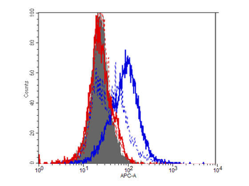
Flow Cytometry of Mouse Anti-IDO1 antibody. Cells: HEK293 cells. Expresing: mouse IDO-1(blue) and mouse IDO-2 (red). Primary antibody: IDO1 (2E2) monoclonal antibody. Secondary antibody: Biotin mouse secondary antibody at 1:10000 for 45 min at RT and streptavidin PE at 1:5000 for 30 min at RT.
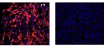
Immunofluorescence Microscopy of Mouse Anti-IDO1 Antibody. Cells: HEK293 cells. Fixation: 0.5% PFA. Expressing: mouse IDO-1 (left) and mouse IDO-2 (right). Primary antibody: IDO1 (2E2) monoclonal antibody. Antigen retrieval: not required. Secondary antibody: mouse secondary antibody at 1:10000 for 45 min at RT. Localization: IDO-1 is located in the cytosol. Staining: IDO1 as red fluorescent signal with bis-benzimide nuclear counterstain (blue).
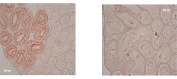
Immunohistochemistry of Mouse Anti-IDO1 Antibody. Tissue: epididymis from wild-type (left) or IDO1 null mice (right). Fixation: frozen sections. Antigen retrieval: not required. Primary antibody: IDO1 (2E2) monoclonal antibody. Secondary antibody: Peroxidase mouse secondary antibody at 1:10000 for 45 min at RT. Localization: IDO-1 is located in the cytosol. Staining: IDO 1 as precipitated brown signal.
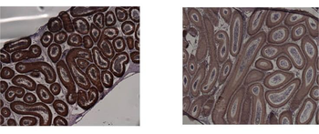
Immunohistochemistry of Mouse anti-IDO1 antibody. Tissue: epididymis from wild-type (left) or IDO1 null mice (right). Fixation: paraffin-embedded. Primary antibody: IDO1 (2E2) monoclonal antibody. Secondary antibody: Peroxidase mouse secondary antibody at 1:10000 for 45 min at RT. Localization: IDO-1 is located in the cytosol. Staining: IDO 1 as precipitated brown signal.

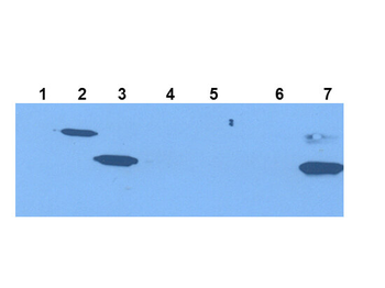
Western Blot of Mouse Anti-IDO1 Antibody. Extracts from 293HEK Cells expressing: Lane 1: Control Vector. Lane 2: His-tagged mouse IDO1. Lane 3: mouse IDO1. Lane 4: His-tagged mouse IDO2. Lane 5: mouse IDO2. Lane 6: Epididymis from IDO null. Lane 7: wild type mice. Primary antibody: IDO-1(2E2) monoclonal antibody. Secondary antibody: IRDye800™ mouse secondary antibody at 1:10000 for 45 min at RT. Block: 1xPBST overnight at 4°C. Predicted/Observed size: 41-42 kDa/41-42 kDa for IDO-1. Other band(s): none.

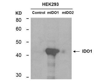
Western Blot of mouse anti-IDO1 antibody. Lane 1: HEK293 control vector. Lane 2: HEK293 expressing mouse IDO1. Lane 3: HEK293 expressing mouse IDO2. Load: 35 µg per lane. Primary antibody: IDO 1 antibody at 1:400 for overnight at 4°C. Secondary antibody: IRDye800™ mouse secondary antibody at 1:10000 for 45 min at RT. Block: 5% BLOTTO overnight at 4°C. Predicted/Observed size: 45.6 kDa, ~44 kDa for IDO1. Other band(s): non-specifics.
- Item 1 of 9
IDO antibody [orb157614]
ICC, IF, IHC-P, WB
Guinea pig, Human, Mouse, Rat
Rabbit
Polyclonal
Unconjugated
100 μg, 200 μg - Item 1 of 5
- Item 1 of 6
IDO antibody [orb345210]
ELISA, FC, IF, IHC, IP, Multiplex Assay, WB
Mouse
Mouse
Monoclonal
Unconjugated
100 μg - Item 1 of 5
- Item 1 of 4
Indoleamine 2, 3-dioxygenase/IDO1 Antibody [orb312129]
FC, ICC, IF, IHC, WB
Human
Rabbit
Polyclonal
Unconjugated
10 μg, 100 μg


Submit a review
Filter by Rating

- 5 stars

- 4 stars
- 3 stars
- 2 stars
- 1 stars
