You have no items in your shopping cart.
Cart summary
Item 1 of 6
Item 1 of 6
IDO antibody
Catalog Number: orb345210

| Catalog Number | orb345210 |
|---|---|
| Category | Antibodies |
| Description | IDO antibody |
| Species/Host | Mouse |
| Clonality | Monoclonal |
| Clone Number | 2E2.6 |
| Tested applications | ELISA, FC, IF, IHC, IP, Multiplex Assay, WB |
| Reactivity | Mouse |
| Isotype | IgG1 |
| Immunogen | IDO1 antibody was produced in mouse by repeated immunizations with mouse recombinant IDO1 protein followed by hybridoma development. |
| Concentration | 1.0 mg/mL |
| Dilution range | ELISA: 1:5000-1:50000, FC: 0.5-1x10^6 cells, IHC: User Optimized, IF: 1:50-1:100, IP: 10-100 µL, WB: 1:500-1:1500 |
| Form/Appearance | Liquid (sterile filtered) |
| Purity | Anti-IDO1 antibody was purified from ascites fluid by Protein A chromatography followed by extensive dialysis against the buffer stated above. IDO1 antibody is specific for mouse IDO1 protein. Mouse IDO1 does not react with human tissues. Cross-reactivity with IDO1 from other sources has not been determined. |
| Conjugation | Unconjugated |
| UniProt ID | P28776 |
| NCBI | NP_032350.1 |
| Storage | Store vial at -20° C prior to opening. Aliquot contents and freeze at -20° C or below for extended storage. Avoid cycles of freezing and thawing. Centrifuge product if not completely clear after standing at room temperature. This product is stable for several weeks at 4° C as an undiluted liquid. Dilute only prior to immediate use. |
| Buffer/Preservatives | 0.01% (w/v) Sodium Azide |
| Alternative names | mouse anti-IDO1 antibody, Ido, Indo, Indoleamine 2 Read more... |
| Note | For research use only |
| Application notes | Anti-IDO1 antibody has been tested for use in ELISA, Western Blot, IF, IHC, and Flow Cytometry. Specific conditions for reactivity should be optimized by the end user. |
| Expiration Date | 12 months from date of receipt. |

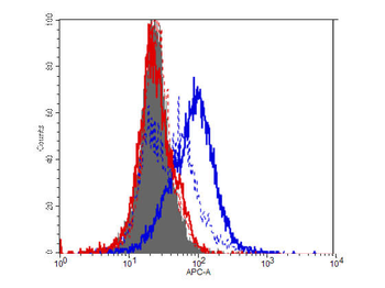
Flow Cytometry of Mouse Anti-IDO1 antibody. Cells: HEK293 cells. Expresing: mouse IDO-1(blue) and mouse IDO-2 (red). Primary antibody: IDO1 (2E2) monoclonal antibody. Secondary antibody: Biotin mouse secondary antibody at 1:10000 for 45 min at RT and streptavidin PE at 1:5000 for 30 min at RT.

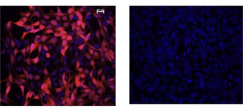
Immunofluorescence Microscopy of Mouse Anti-IDO1 Antibody. Cells: HEK293 cells. Fixation: 0.5% PFA. Expressing: mouse IDO-1 (left) and mouse IDO-2 (right). Primary antibody: IDO1 (2E2) monoclonal antibody. Antigen retrieval: not required. Secondary antibody: mouse secondary antibody at 1:10000 for 45 min at RT. Localization: IDO-1 is located in the cytosol. Staining: IDO1 as red fluorescent signal with bis-benzimide nuclear counterstain (blue).

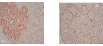
Immunohistochemistry of Mouse Anti-IDO1 Antibody. Tissue: epididymis from wild-type (left) or IDO1 null mice (right). Fixation: frozen sections. Antigen retrieval: not required. Primary antibody: IDO1 (2E2) monoclonal antibody. Secondary antibody: Peroxidase mouse secondary antibody at 1:10000 for 45 min at RT. Localization: IDO-1 is located in the cytosol. Staining: IDO 1 as precipitated brown signal.

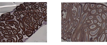
Immunohistochemistry of Mouse anti-IDO1 antibody. Tissue: epididymis from wild-type (left) or IDO1 null mice (right). Fixation: paraffin-embedded. Primary antibody: IDO1 (2E2) monoclonal antibody. Secondary antibody: Peroxidase mouse secondary antibody at 1:10000 for 45 min at RT. Localization: IDO-1 is located in the cytosol. Staining: IDO 1 as precipitated brown signal.
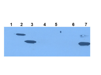
Western Blot of Mouse Anti-IDO1 Antibody. Extracts from 293HEK Cells expressing: Lane 1: Control Vector. Lane 2: His-tagged mouse IDO1. Lane 3: mouse IDO1. Lane 4: His-tagged mouse IDO2. Lane 5: mouse IDO2. Lane 6: Epididymis from IDO null. Lane 7: wild type mice. Primary antibody: IDO-1(2E2) monoclonal antibody. Secondary antibody: IRDye800™ mouse secondary antibody at 1:10000 for 45 min at RT. Block: 1xPBST overnight at 4°C. Predicted/Observed size: 41-42 kDa/41-42 kDa for IDO-1. Other band(s): none.
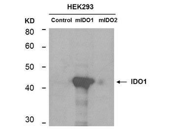
Western Blot of mouse anti-IDO1 antibody. Lane 1: HEK293 control vector. Lane 2: HEK293 expressing mouse IDO1. Lane 3: HEK293 expressing mouse IDO2. Load: 35 µg per lane. Primary antibody: IDO 1 antibody at 1:400 for overnight at 4°C. Secondary antibody: IRDye800™ mouse secondary antibody at 1:10000 for 45 min at RT. Block: 5% BLOTTO overnight at 4°C. Predicted/Observed size: 45.6 kDa, ~44 kDa for IDO1. Other band(s): non-specifics.
- Item 1 of 9
IDO antibody [orb157614]
ICC, IF, IHC-P, WB
Guinea pig, Human, Mouse, Rat
Rabbit
Polyclonal
Unconjugated
100 μg, 200 μg - Item 1 of 5
- Item 1 of 6
IDO antibody [orb345211]
ELISA, FC, IF, IHC, IP, Multiplex Assay, WB
Mouse
Mouse
Monoclonal
Unconjugated
25 μl - Item 1 of 5
- Item 1 of 4
Indoleamine 2, 3-dioxygenase/IDO1 Antibody [orb312129]
FC, ICC, IF, IHC, WB
Human
Rabbit
Polyclonal
Unconjugated
10 μg, 100 μg


Submit a review
Filter by Rating

- 5 stars

- 4 stars
- 3 stars
- 2 stars
- 1 stars
