You have no items in your shopping cart.
Cart summary
Item 1 of 6
Item 1 of 6
ID1 Antibody (Center)
Catalog Number: orb1937995

| Catalog Number | orb1937995 |
|---|---|
| Category | Antibodies |
| Description | Affinity Purified Rabbit Polyclonal Antibody (Pab) |
| Species/Host | Rabbit |
| Clonality | Polyclonal |
| Clone Number | RB29169 |
| Tested applications | FC, IHC-P, WB |
| Predicted Reactivity | Mouse |
| Reactivity | Human, Rat |
| Isotype | Rabbit IgG |
| Antibody Type | Primary Antibody |
| Dilution range | WB: 1:1000, WB: 1:1000, WB: 1:1000, WB: 1:1000, IHC-P: 1:100, FC: 1:25 |
| Form/Appearance | Purified polyclonal antibody supplied in PBS with 0.09% (W/V) sodium azide. This antibody is purified through a protein A column, followed by peptide affinity purification. |
| Conjugation | Unconjugated |
| MW | 16133 Da |
| Target | This ID1 antibody is generated from rabbits immunized with a KLH conjugated synthetic peptide between 66-93 amino acids of human ID1. |
| UniProt ID | P41134 |
| NCBI | NP_851998.1, NP_002156.2 |
| Storage | Maintain refrigerated at 2-8°C for up to 2 weeks. For long term storage store at -20°C in small aliquots to prevent freeze-thaw cycles |
| Alternative names | DNA-binding protein inhibitor ID-1, Class B basic Read more... |
| Note | For research use only |
| Expiration Date | 12 months from date of receipt. |

Anti-ID1 Antibody (Center) at 1:1000 dilution + HepG2 whole cell lysate. Lysates/proteins at 20 µg per lane. Secondary Goat Anti-Rabbit IgG, (H+L), Peroxidase conjugated at 1/10000 dilution. Predicted band size: 16 kDa. Blocking/Dilution buffer: 5% NFDM/TBST.
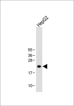
Anti-ID1 Antibody (Center) at 1:1000 dilution + HepG2 whole cell lysate.Lysates/proteins at 20 µg per lane. Secondary Goat Anti-Rabbit IgG, (H+L), Peroxidase conjugated at 1/10000 dilution. Predicted band size: 16 kDa. Blocking/Dilution buffer: 5% NFDM/TBST.
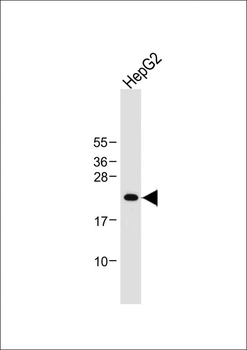
Anti-ID1 Antibody (Center) at 1:1000 dilution + HepG2 whole cell lysate.Lysates/proteins at 20 µg per lane. Secondary Goat Anti-Rabbit IgG, (H+L), Peroxidase conjugated at 1/10000 dilution. Predicted band size: 16 kDa. Blocking/Dilution buffer: 5% NFDM/TBST.
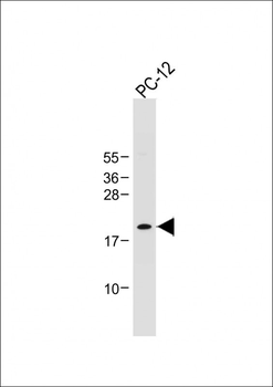
Anti-ID1 Antibody (Center) at 1:1000 dilution + PC-12 whole cell lysate.Lysates/proteins at 20 µg per lane. Secondary Goat Anti-Rabbit IgG, (H+L), Peroxidase conjugated at 1/10000 dilution. Predicted band size: 16 kDa. Blocking/Dilution buffer: 5% NFDM/TBST.
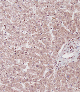
Immunohistochemical analysis on paraffin-embedded Human liver tissue. Tissue was fixed with formaldehyde at room temperature. Heat induced epitope retrieval was performed by EDTA buffer (pH9.0). Samples were incubated with primary antibody (1:100) for 1 hour at room temperature. Undiluted CRF Anti-Polyvalent HRP Polymer antibody was used as the secondary Antibody.
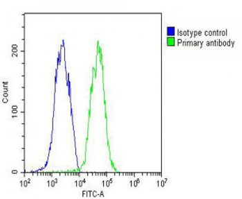
Overlay histogram showing HepG2 cells (green line). The cells were fixed with 2% paraformaldehyde (10 min) and then permeabilized with 90% methanol for 10 min. The cells were then icubated in 2% bovine serum albumin to block non-specific protein-protein interactions followed by the antibody (1:25 dilution) for 60 min at 37°C. The secondary antibody used was Goat-Anti-Rabbit IgG, DyLight 488 Conjugated Highly Cross-Adsorbed at 1/200 dilution for 40 min at 37°C. Isotype control antibody (blue line) was rabbit IgG (1 μg/1x10^6 cells) used under the same conditions. Acquisition of > 10000 events was performed.

ID1 Antibody (Center) [orb1168035]
FC, IF, IHC-P, WB
Human, Rat
Rabbit
Polyclonal
Unconjugated
100 μl, 30 μl
