You have no items in your shopping cart.
Cart summary
Item 1 of 3
Item 1 of 3
ICAM1 Antibody / CD54
Catalog Number: orb2638616

| Catalog Number | orb2638616 |
|---|---|
| Category | Antibodies |
| Description | Recognizes an 85-115kDa protein (variation with cell type), identified as intercellular adhesion molecule (ICAM-1) (Workshop IV). It has 7 potential N-linked glycosylation sites. ICAM-1 is a single chain glycoprotein of Ig supergene family, present on unstimulated endothelial cells (EC) and on a variety of other cell types including activated fibroblasts, EC, macrophages, and lymphocytes. ICAM-1 mediates cell adhesion by binding to integrins CD11a/CD18 (leukocyte adhesion molecule, LFA-1) and to CD11b/CD18 (Mac-1). This interaction enhances antigen-specific T-cell activation. ICAM-1 also binds to CD43 and to Plasmodium falciparum infected RBCs. W-CAM-1 mAb blocks aggregation of cell lines mediated by the ICAM-1 and blocks homotypic binding of purified populations of activated T- and B-lymphocytes and also aggregation of mixed T- and B-cell blasts. It inhibits T-cell adhesion to normal human endothelial cells. Activation induced by cell-cell contact (mixed lymphocyte reaction, T-cell mediated B-cell activation) is significantly inhibited. This mAb blocks elements of both effector arms of immune system (cytotoxic cell function and Ig production). |
| Species/Host | Mouse |
| Clonality | Monoclonal |
| Clone Number | 1H4 or W-CAM-1 or Wehi-CAM-1 |
| Tested applications | FA, FACS, IHC-P |
| Reactivity | Human |
| Isotype | Mouse IgG2b, kappa |
| Immunogen | Raji Burkitt lymphoma cells were used as the immunogen for the ICAM-1 antibody. |
| Antibody Type | Primary Antibody |
| Dilution range | Flow cytometry: 0.5-1ug/million cells in 0.1ml,Functional testing (order BSA/sodium azide-free format),Immunohistochemistry (FFPE): 2-4ug/ml for 30 min at RT (1),Prediluted IHC only format: incubate for 30 min at RT (2) |
| Purity | Protein G affinity chromatography |
| Conjugation | Unconjugated |
| Formula | 0.2 mg/ml in 1X PBS with 0.1 mg/ml BSA (US sourced) and 0.05% sodium azide |
| Hazard Information | This ICAM-1 antibody is available for research use only. |
| UniProt ID | P05362 |
| Storage | Maintain refrigerated at 2-8°C for up to 2 weeks. For long term storage store at -20°C in small aliquots to prevent freeze-thaw cycles. |
| Buffer/Preservatives | 0.2 mg/ml in 1X PBS with 0.1 mg/ml rAlbumin (US sourced) and 0.05% sodium azide |
| Note | For research use only |
| Application notes | Optimal dilution of the ICAM-1 antibody should be determined by the researcher.1. Staining of formalin-fixed tissues requires boiling tissue sections in 10mM Tris with 1mM EDTA, pH 9.0 for 10-20 min followed by cooling at RT for 20 min2. The prediluted format is supplied in a dropper bottle and is optimized for use in IHC. After epitope retrieval step (if required), drip mAb solution onto the tissue section and incubate at RT for 30 min. |
| Expiration Date | 12 months from date of receipt. |

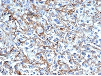
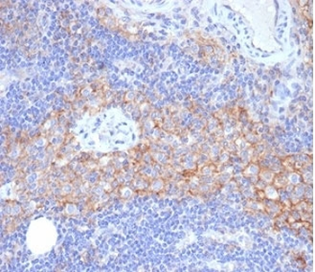
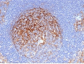
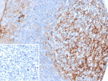
IHC analysis of formalin-fixed, paraffin-embedded human melanoma stained with ICAM-1 antibody (clone 1H4).
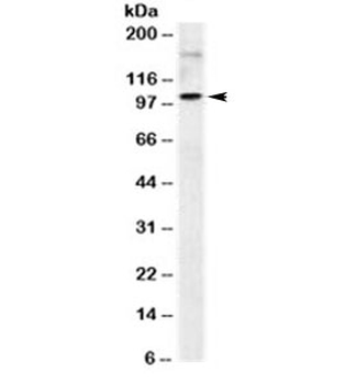
Western blot testing of human Raji cell lysate with ICAM-1 antibody (clone 1H4). Predicted molecular weight: ~58/75-115kDa (unmodified/glycosylated).
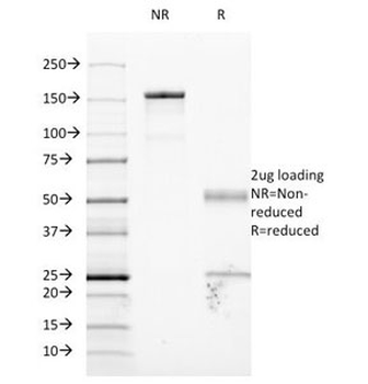
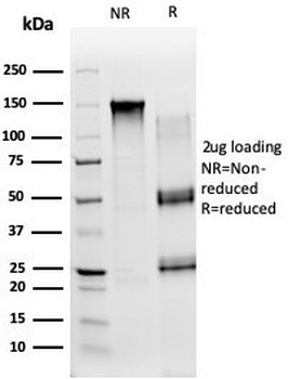
SDS-PAGE Analysis of Purified, BSA-Free ICAM-1 Antibody (clone 1H4 or W-CAM-1 or Wehi-CAM-1). Confirmation of Integrity and Purity of the Antibody.
- Item 1 of 4
- Item 1 of 4
- Item 1 of 3
- Item 1 of 2
- Item 1 of 2