You have no items in your shopping cart.
Cart summary
Item 1 of 6
Item 1 of 6
HSPA5 Antibody
Catalog Number: orb1437589

| Catalog Number | orb1437589 |
|---|---|
| Category | Antibodies |
| Description | Rabbit polyclonal antibody to HSPA5. |
| Species/Host | Rabbit |
| Clonality | Polyclonal |
| Clone Number | RB1768 |
| Tested applications | FC, IF, IHC-P, WB |
| Reactivity | Human, Mouse |
| Isotype | Rabbit IgG |
| Immunogen | Recombinant Protein |
| Dilution range | IF: 1:10~50, WB: 1:1000, WB: 1:1000, IHC-P: 1:50~100, IHC-P: 1:50~100, FC: 1:10~50 |
| Form/Appearance | Purified polyclonal antibody supplied in PBS with 0.09% (W/V) sodium azide. This antibody is prepared by Saturated Ammonium Sulfate (SAS) precipitation followed by dialysis against PBS. |
| Conjugation | Unconjugated |
| MW | 72333 |
| Target | HSPA5 |
| UniProt ID | P11021 |
| NCBI | NP_005338.1 |
| Storage | Maintain refrigerated at 2-8°C for up to 2 weeks. For long term storage store at -20°C in small aliquots to prevent freeze-thaw cycles |
| Alternative names | 78 kDa glucose-regulated protein, GRP-78, Endoplas Read more... |
| Note | For research use only |
| Expiration Date | 12 months from date of receipt. |

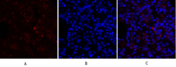
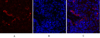
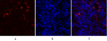
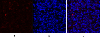
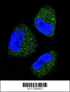
Confocal immunofluorescent analysis of HSPA5 Antibody with NCI-H460 cell followed by Alexa Fluor 488-conjugated goat anti-rabbit lgG (green). DAPI was used to stain the cell nuclear (blue).
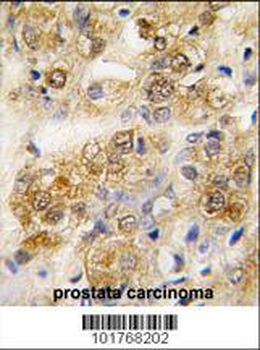
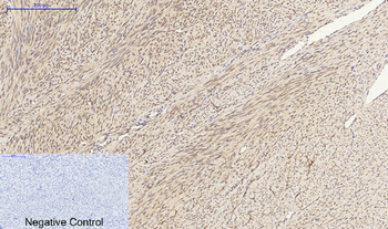
Formalin-fixed and paraffin-embedded human prostata carcinoma tissue reacted with HSPA5 antibody, which was peroxidase-conjugated to the secondary antibody, followed by DAB staining. This data demonstrates the use of this antibody for immunohistochemistry; clinical relevance has not been evaluated.

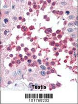
Formalin-fixed and paraffin-embedded human Testis tissue reacted with HSPA5 antibody, which was peroxidase-conjugated to the secondary antibody, followed by AEC staining. This data demonstrates the use of this antibody for immunohistochemistry; clinical relevance has not been evaluated.
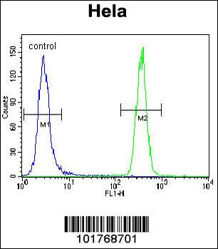
HSPA5 Antibody flow cytometric analysis of Hela cells (right histogram) compared to a negative control cell (left histogram). FITC-conjugated goat-anti-rabbit secondary antibodies were used for the analysis.
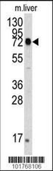
The anti-HSPA5 Pab is used in Western blot to detect HSPA5 in mouse liver tissue lysate. HSPA5 (arrow) was detected using the purified Pab.

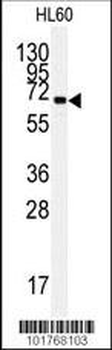
Western blot analysis of anti-HSPA5 Pab in HL60 cell line lysates (35 ug/lane).HSPA5 (arrow) was detected using the purified Pab.
- Item 1 of 8
- Item 1 of 7
HSP A5 Polyclonal Antibody [orb1413406]
IF, IHC-P, WB
Human, Mouse, Rat
Rabbit
Polyclonal
Unconjugated
100 μl - Item 1 of 5
- Item 1 of 5
HSPA5 Antibody [orb676526]
ELISA, IHC, WB
Human, Mouse, Rat
Rabbit
Polyclonal
Unconjugated
50 μg, 100 μg - Item 1 of 6