You have no items in your shopping cart.
Cart summary
Item 1 of 5
Item 1 of 5
HSP90 (total) Antibody
Catalog Number: orb1822480

| Catalog Number | orb1822480 |
|---|---|
| Category | Antibodies |
| Description | Mouse Anti-Human HSP90 (total) Monoclonal IgG2a |
| Species/Host | Mouse |
| Clonality | Monoclonal |
| Clone Number | 4F3.E8 |
| Tested applications | ELISA, ICC, IF, IHC, IP, WB |
| Reactivity | Human, Mouse, Plant, Rat |
| Isotype | IgG2a |
| Immunogen | Recombinant Human HSP90 purified from E.coli |
| Concentration | 1 mg/ml |
| Dilution range | WB (1:2000), IHC (1:100), ICC/IF (1:100); optimal dilutions for assays should be determined by the user. |
| Conjugation | Unconjugated |
| MW | 90kDa |
| Target | HSP90 |
| Entrez | 3320 |
| UniProt ID | P07900 |
| NCBI | NP_001017963.2 |
| Storage | Maintain refrigerated at 2-8°C for up to 2 weeks. For long term storage store at -20°C in small aliquots to prevent freeze-thaw cycles. |
| Buffer/Preservatives | PBS pH 7.2, 50% glycerol, 0.09% sodium azide *Storage buffer changes when conjugated |
| Alternative names | HSP84 Antibody, HSP86 Antibody, HSP90A Antibody, H Read more... |
| Note | For research use only |
| Application notes | 0.5 µg/ml of SMC-149 was sufficient for detection of HSP90alpha in 20 µg of heat shocked HeLa cell lysate by colorimetric immunoblot analysis using Goat anti-mouse IgG:HRP as the secondary antibody. |
| Expiration Date | 12 months from date of receipt. |

Immunocytochemistry/Immunofluorescence analysis using Mouse Anti-Hsp90 Monoclonal Antibody, Clone 4F3.E8. Tissue: HaCaT cells. Species: Human. Fixation: Cold 100% methanol for 10 minutes at -20°C. Primary Antibody: Mouse Anti-Hsp90 Monoclonal Antibody at 1:100 for 1 hour at RT. Secondary Antibody: FITC Goat Anti-Mouse (green) at 1:50 for 1 hour at RT.

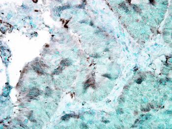
Immunohistochemistry analysis using Mouse Anti-Hsp90 Monoclonal Antibody, Clone 4F3.E8. Tissue: colon carcinoma. Species: Human. Fixation: Formalin. Primary Antibody: Mouse Anti-Hsp90 Monoclonal Antibody at 1:100000 for 12 hours at 4°C. Secondary Antibody: Biotin Goat Anti-Mouse at 1:2000 for 1 hour at RT. Counterstain: Mayer Hematoxylin (purple/blue) nuclear stain at 200 μl for 2 minutes at RT. Localization: Inflammatory cells. Magnification: 40x.

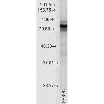
Western Blot analysis of Rat tissue lysate showing detection of Hsp90 protein using Mouse Anti-Hsp90 Monoclonal Antibody, Clone 4F3.E8. Load: 15 μg. Block: 1.5% BSA for 30 minutes at RT. Primary Antibody: Mouse Anti-Hsp90 Monoclonal Antibody at 1:1000 for 2 hours at RT. Secondary Antibody: Sheep Anti-Mouse IgG: HRP for 1 hour at RT.

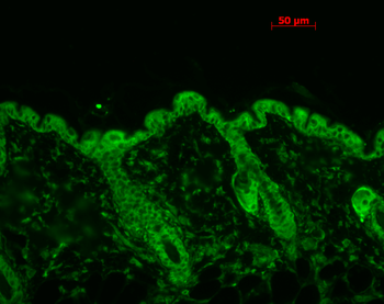
Immunohistochemistry analysis using Mouse Anti-Hsp90 Monoclonal Antibody, Clone 4F3.E8. Tissue: backskin. Species: Mouse. Fixation: Bouin's Fixative and paraffin-embedded. Primary Antibody: Mouse Anti-Hsp90 Monoclonal Antibody at 1:100 for 1 hour at RT. Secondary Antibody: FITC Goat Anti-Mouse (green) at 1:50 for 1 hour at RT.

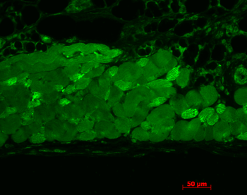
Immunohistochemistry analysis using Mouse Anti-Hsp90 Monoclonal Antibody, Clone 4F3.E8. Tissue: muscle tissue. Species: Mouse. Fixation: Bouin's Fixative and paraffin-embedded. Primary Antibody: Mouse Anti-Hsp90 Monoclonal Antibody at 1:1000 for 1 hour at RT. Secondary Antibody: FITC Goat Anti-Mouse (green) at 1:50 for 1 hour at RT.
- Item 1 of 5
HSP90 (total) Antibody: APC [orb147312]
ICC, IF, IHC
Human, Mouse, Plant, Rat
Mouse
Monoclonal
APC
200 μg - Item 1 of 5
HSP90 (total) Antibody: Biotin [orb147313]
ELISA, ICC, IF, IHC, WB
Human, Mouse, Plant, Rat
Mouse
Monoclonal
Biotin
200 μg - Item 1 of 5
HSP90 (total) Antibody: FITC [orb147314]
ICC, IF, IHC
Human, Mouse, Plant, Rat
Mouse
Monoclonal
FITC
200 μg - Item 1 of 5
HSP90 (total) Antibody: HRP [orb147315]
ELISA, IHC, WB
Human, Mouse, Plant, Rat
Mouse
Monoclonal
HRP
200 μg - Item 1 of 5
HSP90 (total) Antibody: PerCP [orb147317]
ELISA, ICC, IF, IHC, WB
Human, Mouse, Plant, Rat
Mouse
Monoclonal
PerCP
200 μg