You have no items in your shopping cart.
Cart summary
Item 1 of 8
Item 1 of 8
HSP90 Antibody: RPE
Catalog Number: orb146808

| Catalog Number | orb146808 |
|---|---|
| Category | Antibodies |
| Description | Mouse monoclonal to Hsp90 (RPE). HSP90 is an abundantly and ubiquitously expressed heat... |
| Species/Host | Mouse |
| Clonality | Monoclonal |
| Clone Number | H9010 |
| Tested applications | ICC, IF, IHC, WB |
| Reactivity | Canine, Fish, Gallus, Hamster, Human, Mouse, Rabbit, Rat |
| Isotype | IgG2a |
| Immunogen | Recombinant human HSP90beta |
| Concentration | 1 mg/ml |
| Dilution range | WB (1:2500), IHC (1:100) |
| Conjugation | RPE |
| MW | 90kDa |
| Target | HSP90 |
| Entrez | 3326 |
| UniProt ID | P08238 |
| NCBI | NP_031381.2 |
| Storage | Conjugated antibodies should be stored according to the product label |
| Buffer/Preservatives | 95.64mM Phosphate, 2.48mM MES and 2mM EDTA |
| Alternative names | HSP84 antibody, HSP90 antibody, HSP90 beta antibod Read more... |
| Note | For research use only |
| Application notes | 1 µg/ml of SMC-107 was sufficient for detection of HSP90beta in 20 µg of heat shocked HeLa cell lysate by colorimetric immunoblot analysis using Goat anti-mouse IgG:HRP as the secondary antibody. |
| Expiration Date | 12 months from date of receipt. |

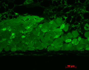
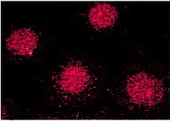
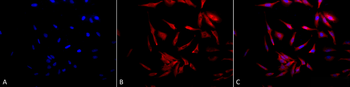
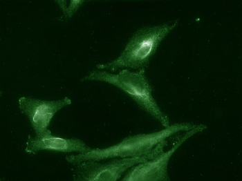
Immunohistochemistry analysis using Mouse Anti-Hsp90 Monoclonal Antibody, Clone H9010. Tissue: inflamed colon. Species: Mouse. Fixation: Formalin. Primary Antibody: Mouse Anti-Hsp90 Monoclonal Antibody at 1:10000 for 12 hours at 4°C. Secondary Antibody: Alexa Fluor 555 Goat Anti-Mouse (red) at 1:5000 for 1 hour at RT. Localization: Inflammatory and epithelial mucosa. Magnification: 40x. Inflammatory and epithelial mucosa.
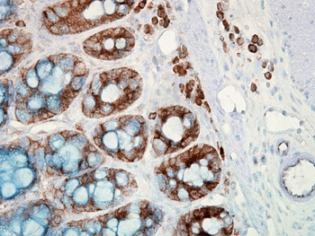
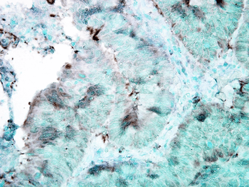
Immunohistochemistry analysis using Mouse Anti-Hsp90 Monoclonal Antibody, Clone H9010. Tissue: colon carcinoma. Species: Human. Fixation: Formalin. Primary Antibody: Mouse Anti-Hsp90 Monoclonal Antibody at 1:10000 for 12 hours at 4°C. Secondary Antibody: Biotin Goat Anti-Mouse at 1:2000 for 1 hour at RT. Counterstain: Mayer Hematoxylin (purple/blue) nuclear stain at 200 μl for 2 minutes at RT. Localization: Inflammatory cells. Magnification: 40x.

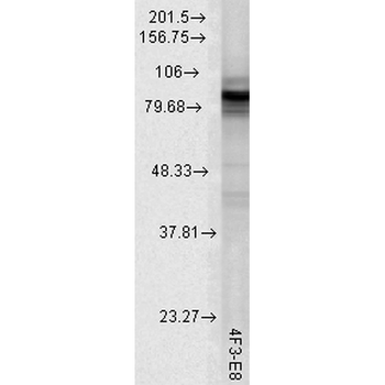
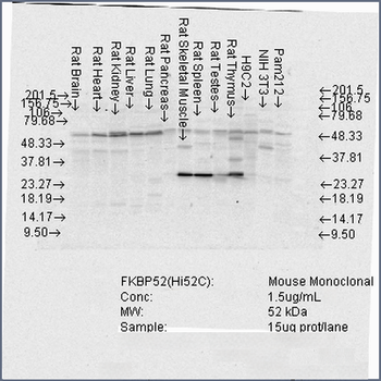
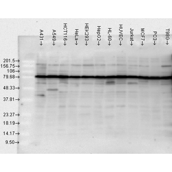
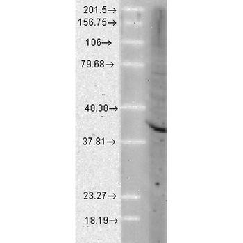
Western Blot analysis of Human cell lysates from various cell lines showing detection of Hsp90 protein using Mouse Anti-Hsp90 Monoclonal Antibody, Clone H9010. Load: 15 μg. Block: 1.5% BSA for 30 minutes at RT. Primary Antibody: Mouse Anti-Hsp90 Monoclonal Antibody at 1:1000 for 2 hours at RT. Secondary Antibody: Sheep Anti-Mouse IgG: HRP for 1 hour at RT.
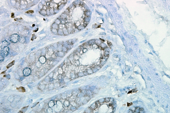
Immunohistochemistry analysis using Mouse Anti-Hsp90 Monoclonal Antibody, Clone H9010. Tissue: inflamed colon. Species: Mouse. Fixation: Formalin. Primary Antibody: Mouse Anti-Hsp90 Monoclonal Antibody at 1:10000 for 12 hours at 4°C. Secondary Antibody: Biotin Goat Anti-Mouse at 1:2000 for 1 hour at RT. Counterstain: Mayer Hematoxylin (purple/blue) nuclear stain at 200 μl for 2 minutes at RT. Localization: Inflammatory cells. Magnification: 40x.
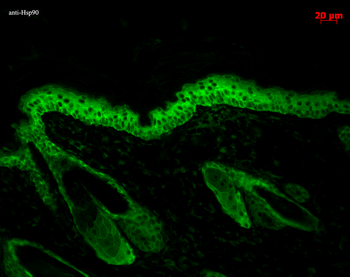
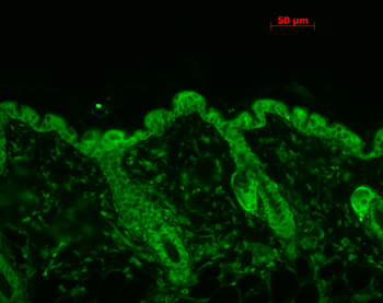
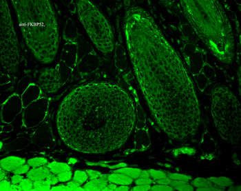
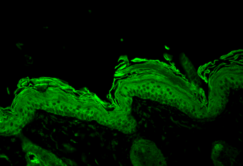
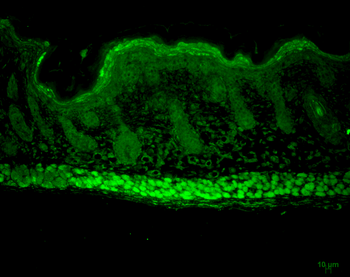
Immunohistochemistry analysis using Mouse Anti-Hsp90 Monoclonal Antibody, Clone H9010. Tissue: backskin. Species: Mouse. Fixation: Bouin's Fixative and paraffin-embedded. Primary Antibody: Mouse Anti-Hsp90 Monoclonal Antibody at 1:100 for 1 hour at RT. Secondary Antibody: FITC Goat Anti-Mouse (green) at 1:50 for 1 hour at RT. Localization: Epidermis.
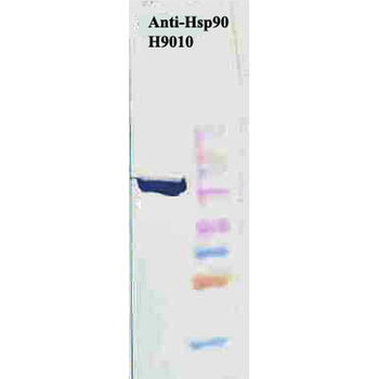
Western Blot analysis of Human Cervical cancer cell line (HeLa) lysate showing detection of Hsp90 protein using Mouse Anti-Hsp90 Monoclonal Antibody, Clone H9010. Primary Antibody: Mouse Anti-Hsp90 Monoclonal Antibody at 1:1000. Secondary Antibody: HRP Goat Anti-Mouse.
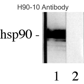
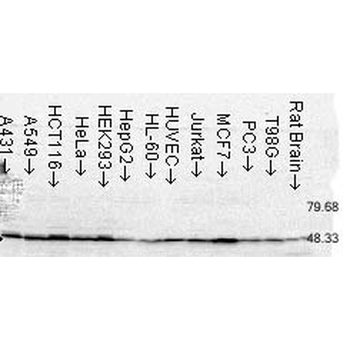
Western blot analysis of Human Lysates showing detection of Hsp90 protein using Mouse Anti-Hsp90 Monoclonal Antibody, Clone H9010. Primary Antibody: Mouse Anti-Hsp90 Monoclonal Antibody at 1:1000. Comparison of clone H9010 behavior with Hsp90 human beta (1) and Hsp90 human alpha (2).
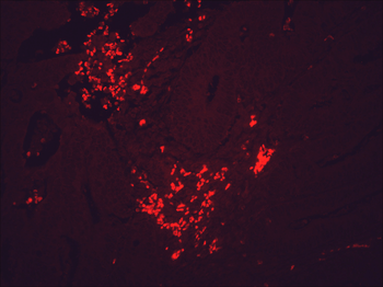
Immunohistochemistry analysis using Mouse Anti-Hsp90 Monoclonal Antibody, Clone H9010. Tissue: colon carcinoma. Species: Human. Fixation: Formalin. Primary Antibody: Mouse Anti-Hsp90 Monoclonal Antibody at 1:10000 for 12 hours at 4°C. Secondary Antibody: Alexa Fluor 555 Goat Anti-Mouse (red) at 1:5000 for 1 hour at RT. Magnification: 40x.
- Item 1 of 5
HSP90 (total) Antibody: RPE [orb147318]
ICC, IF, IHC, WB
Human, Mouse, Plant, Rat
Mouse
Monoclonal
RPE
200 μg - Item 1 of 5
FKBP52 Antibody: RPE [orb147216]
ICC, IF, IHC, WB
Canine, Hamster, Human, Mouse, Rat
Mouse
Monoclonal
RPE
100 μg - Item 1 of 4
- Item 1 of 4
- Item 1 of 4