You have no items in your shopping cart.
Cart summary
Item 1 of 5
Item 1 of 5
HSP70 Antibody: APC
Catalog Number: orb146735

| Catalog Number | orb146735 |
|---|---|
| Category | Antibodies |
| Description | Mouse monoclonal to Hsp70 (APC). Hsp70 genes encode abundant heat-inducible 70-kDa hsps (hsp70s). In most eukaryotes hsp70 genes exist as part of a multigene family. They are found in most cellular compartments of eukaryotes including nuclei, mitochondria, chloroplasts, the endoplasmic reticulum and the cytosol, as well as in bacteria. The genes show a high degree of conservation, having at least 5O% identity. The N-terminal two thirds of hsp70s are more conserved than the C-terminal third. Hsp70 binds ATP with high affinity and possesses a weak ATPase activity which can be stimulated by binding to unfolded proteins and synthetic peptides. When hsc70 (constitutively expressed) present in mammalian cells was truncated, ATP binding activity was found to reside in an N-terminal fragment of 44 kDa which lacked peptide binding capacity. Polypeptide binding ability therefore resided within the C-terminal half. The structure of this ATP binding domain displays multiple features of nucleotide binding proteins. All hsp70s, regardless of location, bind proteins, particularly unfolded ones. The molecular chaperones of the hsp70 family recognize and bind to nascent polypeptide chains as well as partially folded intermediates of proteins preventing their aggregation and misfolding. The binding of ATP triggers a critical conformational change leading to the release of the bound substrate protein. The universal ability of hsp70s to undergo cycles of binding to and release from hydrophobic stretches of partially unfolded proteins determines their role in a great variety of vital intracellular functions such as protein synthesis, protein folding and oligomerization and protein transport. |
| Species/Host | Mouse |
| Clonality | Monoclonal |
| Clone Number | C92F3A-5 |
| Tested applications | ICC, IF, IHC |
| Reactivity | Bovine, Canine, Drosophila, Fish, Gallus, Guinea pig, Hamster, Human, Monkey, Mouse, Other, Porcine, Rabbit, Rat, Sheep |
| Isotype | IgG1 |
| Immunogen | Human HSP70 |
| Concentration | 1 mg/ml |
| Dilution range | WB (1:1000), IHC (1:10000), ICC/IF (1:1000), FACS (1:1000) |
| Conjugation | APC |
| MW | 70kDa |
| Target | HSP70 |
| Entrez | 3303 |
| UniProt ID | P0DMV9, P0DMV8 |
| NCBI | NP_005336.3 |
| Storage | Conjugated antibodies should be stored according to the product label |
| Buffer/Preservatives | 95.64mM Phosphate, 2.48mM MES and 2mM EDTA |
| Alternative names | HSP70 1 antibody, HSP70 2 antibody, HSP70.1 antibo Read more... |
| Note | For research use only |
| Application notes | 1 µg/ml of SMC-100 was sufficient for detection of HSP70 in 20 µg of heat shocked HeLa cell lysate by colorimetric immunoblot analysis using Goat anti-mouse IgG:HRP as the secondary antibody. |
| Expiration Date | 12 months from date of receipt. |

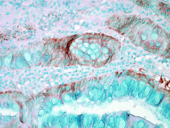
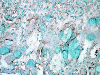
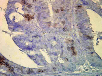
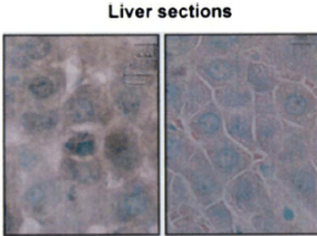
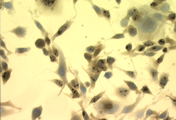
Immunohistochemistry analysis using Mouse Anti-Hsp70 Monoclonal Antibody, Clone C92F3A-5. Tissue: colon carcinoma. Species: Mouse. Fixation: Formalin. Primary Antibody: Mouse Anti-Hsp70 Monoclonal Antibody at 1:10000 for 12 hours at 4°C. Secondary Antibody: Biotin Goat Anti-Mouse at 1:2000 for 1 hour at RT. Counterstain: Mayer Hematoxylin (purple/blue) nuclear stain at 200 μl for 2 minutes at RT. Localization: Inflammatory cells. Magnification: 40x.
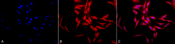
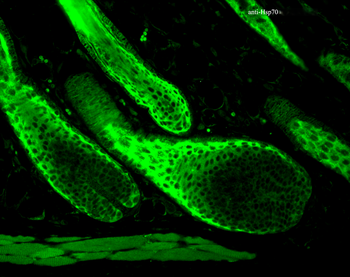
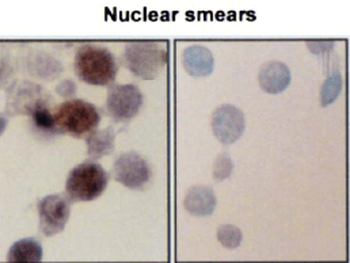
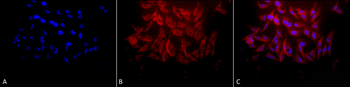
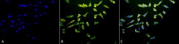
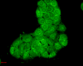
Immunocytochemistry/Immunofluorescence analysis using Mouse Anti-Hsp70 Monoclonal Antibody, Clone C92F3A-5. Tissue: Heat Shocked Melanoma cells. Species: Mouse. Fixation: Formalin. Primary Antibody: Mouse Anti-Hsp70 Monoclonal Antibody at 1:1000 for 16 hours at RT. Secondary Antibody: Biotin Goat Anti-Mouse.
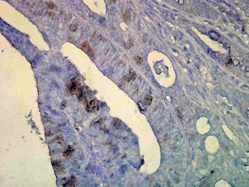
Immunohistochemistry analysis using Mouse Anti-Hsp70 Monoclonal Antibody, Clone C92F3A-5. Tissue: colon carcinoma. Species: Human. Fixation: Formalin. Primary Antibody: Mouse Anti-Hsp70 Monoclonal Antibody at 1:10000 for 12 hours at 4°C. Secondary Antibody: Biotin Goat Anti-Mouse at 1:2000 for 1 hour at RT. Counterstain: Mayer Hematoxylin (purple/blue) nuclear stain at 200 μl for 2 minutes at RT. Localization: Inflammatory cells. Magnification: 40x.

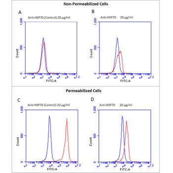
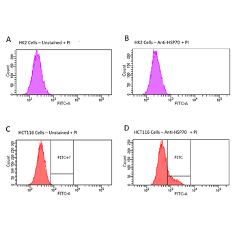
Fluorescence Activated Cell Sorting analysis using Mouse Anti-Hsp70: FITC Monoclonal Antibody, Clone C92F3A-5. Tissue: Heat Shocked CD3+ CD8+ T cells. Species: Mouse. Primary Antibody: Mouse Anti-Hsp70: FITC Monoclonal Antibody at 1:1000.
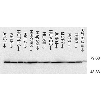
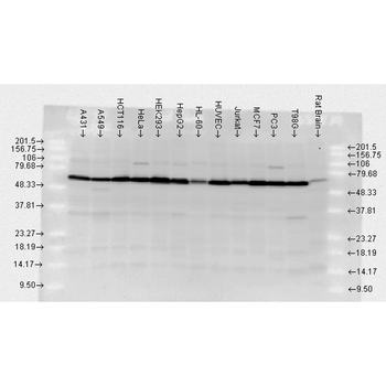
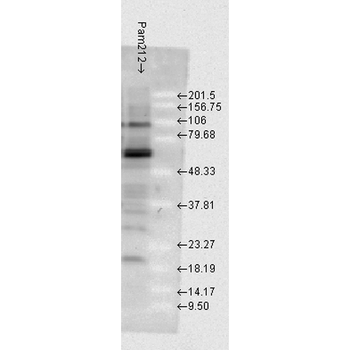
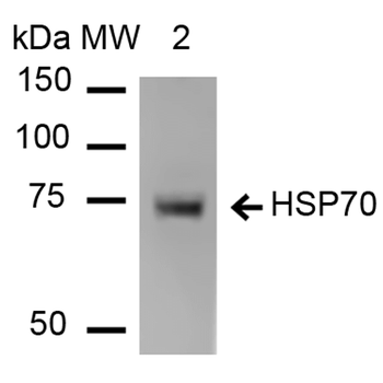
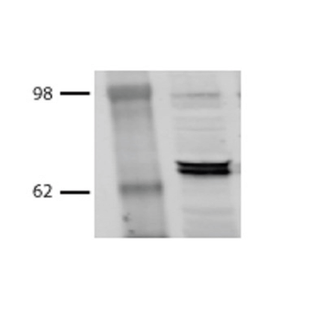
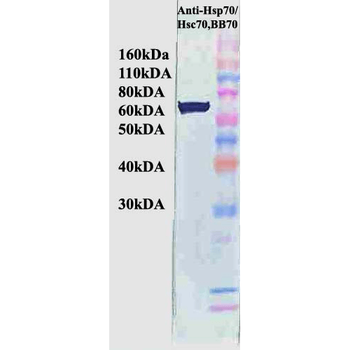
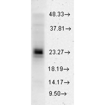
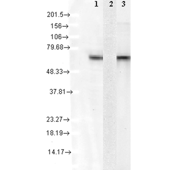
Western Blot analysis of Human cell lysates from various cell lines showing detection of Hsp70 protein using Mouse Anti-Hsp70 Monoclonal Antibody, Clone C92F3A-5. Load: 15 μg. Block: 1.5% BSA for 30 minutes at RT. Primary Antibody: Mouse Anti-Hsp70 Monoclonal Antibody at 1:1000 for 2 hours at RT. Secondary Antibody: Sheep Anti-Mouse IgG: HRP for 1 hour at RT.
- Item 1 of 7
HSP70 Antibody: APC [orb151240]
ICC, IF, IHC
Bovine, Canine, Fish, Guinea pig, Hamster, Human, Mammal, Monkey, Mouse, Other, Plant, Porcine, Rat, Sheep
Rabbit
Polyclonal
APC
100 μg - Item 1 of 6
HSP70 Antibody: APC [orb54777]
ICC, IF, IHC
Bovine, Human, Mouse, Porcine, Rat
Mouse
Monoclonal
APC
100 μg - Item 1 of 6
HSP70/HSC70 Antibody: APC [orb146785]
ICC, IF, IHC
Bovine, Canine, Drosophila, Fish, Frog, Gallus, Guinea pig, Hamster, Human, Mouse, Other, Porcine, Rabbit, Rat, Sheep, Yeast
Mouse
Monoclonal
APC
200 μg - Item 1 of 4
- Item 1 of 4