You have no items in your shopping cart.
Cart summary
Item 1 of 4
Item 1 of 4
HSP60 Antibody: PerCP
Catalog Number: orb151279

| Catalog Number | orb151279 |
|---|---|
| Category | Antibodies |
| Description | Rabbit polyclonal to Hsp60 (PerCP). In both prokaryotic and eukaryotic cells, the misfolding In both prokaryotic and eukaryotic cells, the misfolding... |
| Species/Host | Rabbit |
| Clonality | Polyclonal |
| Tested applications | ELISA, ICC, IF, IHC, WB |
| Reactivity | Bovine, Canine, Gallus, Hamster, Human, Mouse, Rabbit, Rat |
| Immunogen | Human HSP60 produced through recombinant DNA methods in E.coli |
| Concentration | 1 mg/ml |
| Dilution range | WB (1:1000), ICC/IF (1:100) |
| Conjugation | PerCP |
| MW | 60kDa |
| Target | HSP60 |
| Entrez | 3329 |
| UniProt ID | P10809 |
| NCBI | NP_002147.2 |
| Storage | Conjugated antibodies should be stored according to the product label |
| Buffer/Preservatives | 95.64mM Phosphate, 2.48mM MES and 2mM EDTA |
| Alternative names | CPN60 antibody, GROEL antibody, HLD4 antibody, HSP Read more... |
| Note | For research use only |
| Application notes | 1 µg/ml of SPC-105 was sufficient for detection of HSP60 in 20 µg of heat shocked HeLa cell lysate by colorimetric immunoblot analysis using goat anti-mouse IgG as the secondary antibody. |
| Expiration Date | 12 months from date of receipt. |

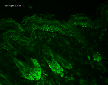
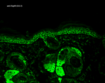
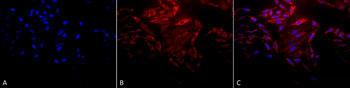
Immunocytochemistry/Immunofluorescence analysis using Rabbit Anti-Hsp60 Polyclonal Antibody. Tissue: Heat Shocked Cervical cancer cell line (HeLa). Species: Human. Fixation: 2% Formaldehyde for 20 min at RT. Primary Antibody: Rabbit Anti-Hsp60 Polyclonal Antibody at 1:100 for 12 hours at 4°C. Secondary Antibody: FITC Goat Anti-Rabbit (green) at 1:200 for 2 hours at RT. Counterstain: DAPI (blue) nuclear stain at 1:40000 for 2 hours at RT. Localization: Mitochondrion matrix. Magnification: 100x. (A) DAPI (blue) nuclear stain. (B) Anti-Hsp60 Antibody. (C) Composite. Heat Shocked at 42°C for 1h.
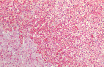
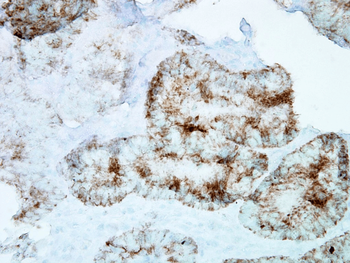
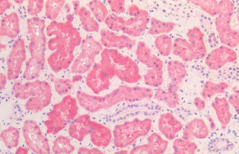
Immunohistochemistry analysis using Rabbit Anti-HSP60 Polyclonal Antibody. Tissue: Adrenal. Species: Human. Fixation: Formalin fixed paraffin-embedded. Primary Antibody: Rabbit Anti-HSP60 Polyclonal Antibody.
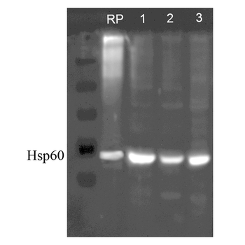
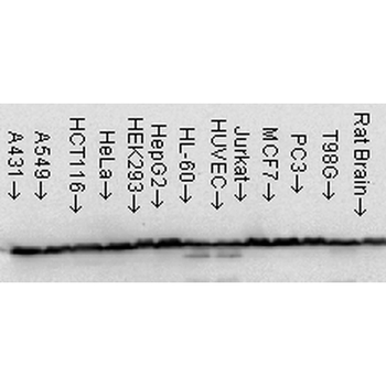
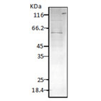
Western blot analysis of Human, Dog, Mouse SKBR3, MDCK, and MEF cell line lysates showing detection of HSP60 protein using Rabbit Anti-HSP60 Polyclonal Antibody. Lane 1: Recom. Human Hsp60 (100ng), Lane2, 3 and 4: SKBR3 lysate (human), MDCK lysate (dog) and MEF lysate (mouse) (al at 7.5ug). Primary Antibody: Rabbit Anti-HSP60 Polyclonal Antibody at 1:1000.
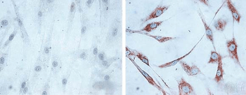
Immunocytochemistry/Immunofluorescence analysis using Rabbit Anti-Hsp60 Polyclonal Antibody. Tissue: Heat Shocked Cervical cancer cell line (HeLa). Species: Human. Fixation: 2% Formaldehyde for 20 min at RT. Primary Antibody: Rabbit Anti-Hsp60 Polyclonal Antibody at 1:100 for 12 hours at 4°C. Secondary Antibody: APC Goat Anti-Rabbit (red) at 1:200 for 2 hours at RT. Counterstain: DAPI (blue) nuclear stain at 1:40000 for 2 hours at RT. Localization: Mitochondrion matrix. Magnification: 20x. (A) DAPI (blue) nuclear stain. (B) Anti-Hsp60 Antibody. (C) Composite. Heat Shocked at 42°C for 1h.
- Item 1 of 5
HSP60 Antibody: PerCP [orb146858]
ELISA, ICC, IF, IHC, WB
Bovine, Canine, Drosophila, Frog, Gallus, Guinea pig, Hamster, Human, Monkey, Mouse, Other, Plant, Porcine, Rabbit, Rat, Sheep
Mouse
Monoclonal
PerCP
200 μg - Item 1 of 4
HSP60 Antibody: PerCP [orb146875]
ELISA, ICC, IF, IHC, WB
Bacteria, Bovine, Canine, E. coli, Fish, Gallus, Guinea pig, H. pylori, Hamster, Human, Insect, Monkey, Mouse, Other, Plant, Porcine, Rabbit, Rat, Yeast
Mouse
Monoclonal
PerCP
200 μg - Item 1 of 2
HSP60 (P. falciparum) Antibody: PerCP [orb152053]
ELISA, ICC, IF, IHC, WB
Bacteria, E. coli
Rabbit
Polyclonal
PerCP
100 μg - Item 1 of 2
Cpn10 Antibody: PerCP [orb152104]
ELISA, ICC, IF, IHC, WB
Bovine, Canine, Frog, Guinea pig, Human, Mouse, Porcine, Rabbit, Rat, Sheep
Rabbit
Polyclonal
PerCP
100 μg 
HSP60 Rabbit Polyclonal Antibody (PerCP-Cy7) [orb1594732]
ICC, IF
Bovine, Canine, Equine, Rabbit
Human, Mouse, Rat
Rabbit
Polyclonal
PerCP/Cy7
100 μl