You have no items in your shopping cart.
Cart summary
Item 1 of 5
Item 1 of 5
Hsp60 Antibody
Catalog Number: orb349729

| Catalog Number | orb349729 |
|---|---|
| Category | Antibodies |
| Description | Rabbit polyclonal antibody to HSPD1. |
| Species/Host | Rabbit |
| Clonality | Polyclonal |
| Clone Number | RB55969 |
| Tested applications | IF, IHC-P, WB |
| Reactivity | Human, Mouse |
| Isotype | Rabbit IgG |
| Immunogen | Synthesized Peptide |
| Dilution range | IF: 1:25, WB: 1:2000, WB: 1:2000, IHC-P: 1:25, IHC-P: 1:25 |
| Form/Appearance | Purified polyclonal antibody supplied in PBS with 0.09% (W/V) sodium azide. This antibody is purified through a protein A column, followed by peptide affinity purification. |
| Conjugation | Unconjugated |
| MW | 61055 |
| Target | HSPD1 |
| UniProt ID | P10809 |
| Storage | Maintain refrigerated at 2-8°C for up to 2 weeks. For long term storage store at -20°C in small aliquots to prevent freeze-thaw cycles |
| Alternative names | 60 kDa heat shock protein, mitochondrial, 60 kDa c Read more... |
| Note | For research use only |
| Expiration Date | 12 months from date of receipt. |

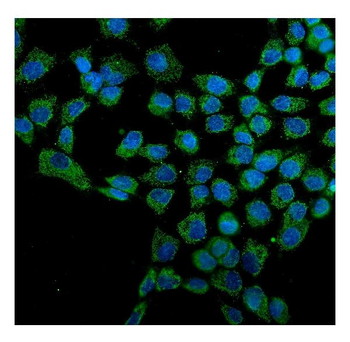
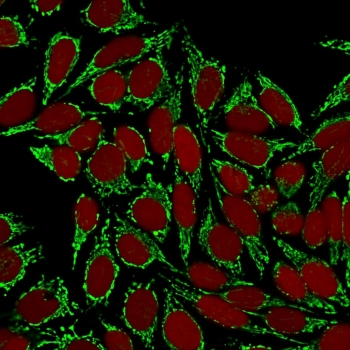
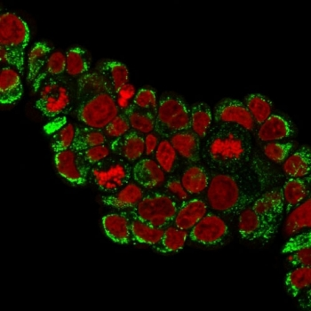
Immunofluorescent analysis of 4% paraformaldehyde-fixed, 0.1% Triton X-100 permeabilized HepG2 (human liver hepatocellular carcinoma cell line) cells labeling Hsp60 at 1/25 dilution, followed by Dylight 488-conjugated goat anti-rabbit IgG secondary antibody at 1/200 dilution (green). Immunofluorescence image showing cytoplasm staining on HepG2 cell line. The nuclear counter stain is DAPI (blue).
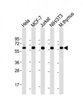
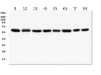
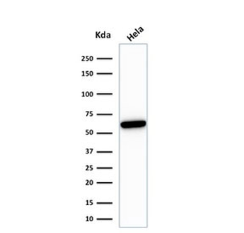
All lanes: Anti-Hsp60 Antibody at 1:2000 dilution. Lane 1: Hela whole cell lysate. Lane 2: MCF-7 whole cell lysate. Lane 3: Jurkat whole cell lysate. Lane 4: NIH/3T3 whole cell lysate. Lane 5: mouse thymus lysate. Lysates/proteins at 20 µg per lane. Secondary Goat Anti-Rabbit IgG, (H+L), Peroxidase conjugated at 1/10000 dilution. Predicted band size: 61 kDa. Blocking/Dilution buffer: 5% NFDM/TBST.
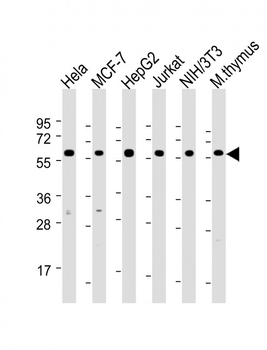
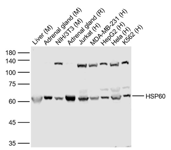
All lanes: Anti-Hsp60 Antibody at 1:2000 dilution. Lane 1: Hela whole cell lysate. Lane 2: MCF-7 whole cell lysate. Lane 3: HepG2 whole cell lysate. Lane 4: Jurkat whole cell lysate. Lane 5: NIH/3T3 whole cell lysate. Lane 6: mouse thymus lysate. Lysates/proteins at 20 µg per lane. Secondary Goat Anti-Rabbit IgG, (H+L), Peroxidase conjugated at 1/10000 dilution. Predicted band size: 61 kDa. Blocking/Dilution buffer: 5% NFDM/TBST.
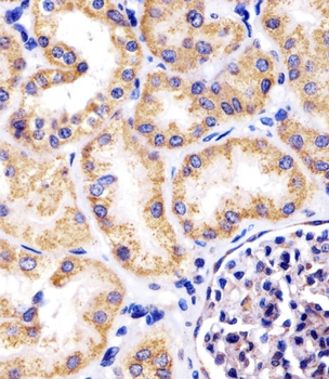
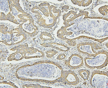
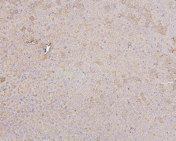
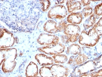
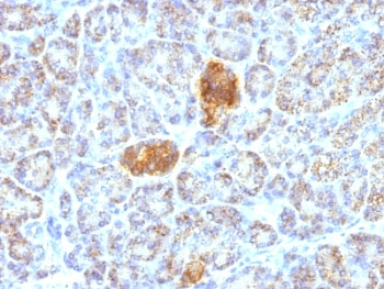
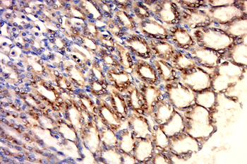
Staining Hsp60 in human kidney tissue sections by Immunohistochemistry (IHC-P - paraformaldehyde-fixed, paraffin-embedded sections). Tissue was fixed with formaldehyde and blocked with 3% BSA for 0.5 hour at room temperature; antigen retrieval was by heat mediation with a citrate buffer (pH6). Samples were incubated with primary antibody (1/25) for 1 hours at 37°C. A undiluted biotinylated goat polyvalent antibody was used as the secondary antibody.
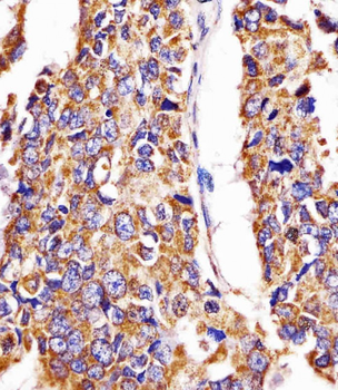
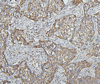
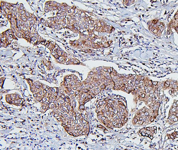
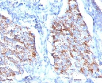
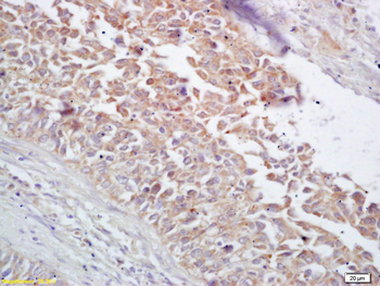
Staining Hsp60 in human lung adenocarcinoma tissue sections by Immunohistochemistry (IHC-P - paraformaldehyde-fixed, paraffin-embedded sections). Tissue was fixed with formaldehyde and blocked with 3% BSA for 0.5 hour at room temperature; antigen retrieval was by heat mediation with a citrate buffer (pH6). Samples were incubated with primary antibody (1/25) for 1 hours at 37°C. A undiluted biotinylated goat polyvalent antibody was used as the secondary antibody.
- Item 1 of 9
Anti-Hsp60/HSPD1 Antibody (monoclonal, 6G2) [orb570314]
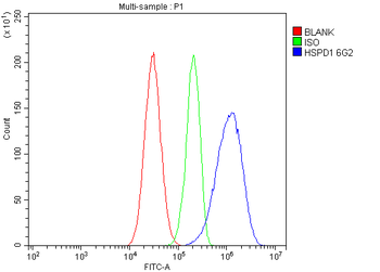
FC, ICC, IF, IHC, WB
Human, Mouse, Rat
Mouse
Monoclonal
Unconjugated
10 μg, 100 μg - Item 1 of 8
- Item 1 of 8
- Item 1 of 5
HSP60 Rabbit Polyclonal Antibody [orb10846]
ICC, IF, IHC-Fr, IHC-P, WB
Bovine, Canine, Equine, Rabbit
Human, Mouse, Rat
Rabbit
Polyclonal
Unconjugated
50 μl, 100 μl, 200 μl - Item 1 of 4
Goat anti-HSP60 / HSPD1 (aa333-344) Antibody [orb334054]
ELISA, IF, IHC, WB
Bovine, Canine, Human, Porcine
Goat
Polyclonal
Unconjugated
100 μg