You have no items in your shopping cart.
Cart summary
Item 1 of 5
Item 1 of 5
HSP60 Antibody: Biotin
Catalog Number: orb146854

| Catalog Number | orb146854 |
|---|---|
| Category | Antibodies |
| Description | Mouse monoclonal to Hsp60 (Biotin). In both prokaryotic and eukaryotic cells, the misfolding In both prokaryotic and eukaryotic cells, the misfolding... |
| Species/Host | Mouse |
| Clonality | Monoclonal |
| Clone Number | LK1 |
| Tested applications | ELISA, ICC, IF, IHC, WB |
| Reactivity | Bovine, Canine, Drosophila, Frog, Gallus, Guinea pig, Hamster, Human, Monkey, Mouse, Other, Plant, Porcine, Rabbit, Rat, Sheep |
| Isotype | IgG1 |
| Immunogen | Recombinant human HSP60 |
| Concentration | 1 mg/ml |
| Dilution range | WB (1:20000), IHC (1:100), ICC/IF (1:100), IP (1:200) |
| Conjugation | Biotin |
| MW | 60kDa |
| Target | HSP60 |
| Entrez | 3329 |
| UniProt ID | P10809 |
| NCBI | NP_002147.2 |
| Storage | Conjugated antibodies should be stored according to the product label |
| Buffer/Preservatives | 136.36mM Ethanolamine, and 9.55mM Sodium Bicarbonate in 95.45% PBS |
| Alternative names | CPN60 antibody, GROEL antibody, HLD4 antibody, HSP Read more... |
| Note | For research use only |
| Application notes | 0.05 µg/ml of SMC-110 was sufficient for detection of HSP60 in 20 µg of heat shocked HeLa cell lysate by colorimetric immunoblot analysis using goat anti-mouse IgG as the secondary antibody. |
| Expiration Date | 12 months from date of receipt. |

Immunocytochemistry/Immunofluorescence analysis using Mouse Anti-Hsp60 Monoclonal Antibody, Clone LK-1. Tissue: HaCaT cells. Species: Human. Fixation: Cold 100% methanol at -20°C for 10 minutes. Primary Antibody: Mouse Anti-Hsp60 Monoclonal Antibody at 1:100 for 1 hour at RT. Secondary Antibody: FITC Goat Anti-Mouse (green) at 1:50 for 1 hour at RT. Localization: Cytoplasmic Staining.
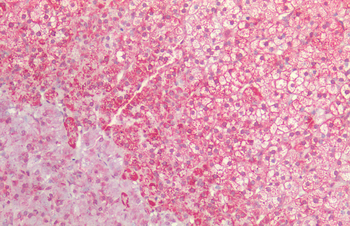
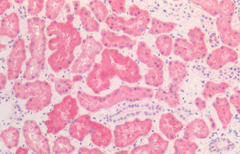
Immunohistochemistry analysis using Mouse Anti-Hsp60 Monoclonal Antibody, Clone LK-1. Tissue: colon carcinoma. Species: Human. Fixation: Formalin. Primary Antibody: Mouse Anti-Hsp60 Monoclonal Antibody at 1:100000 for 12 hours at 4°C. Secondary Antibody: Biotin Goat Anti-Mouse at 1:2000 for 1 hour at RT. Counterstain: Mayer Hematoxylin (purple/blue) nuclear stain at 200 μl for 2 minutes at RT. Localization: Inflammatory cells. Magnification: 40x.
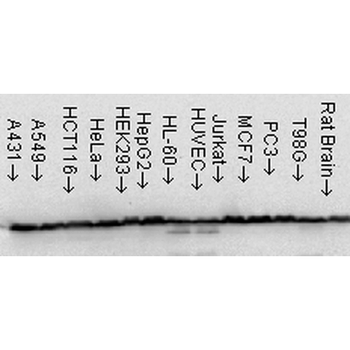
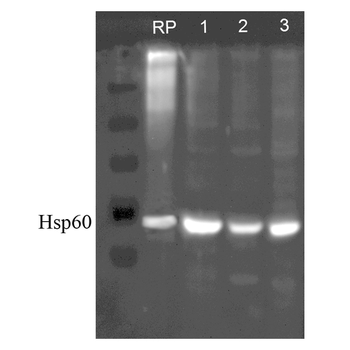
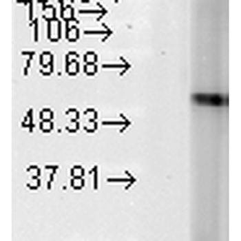
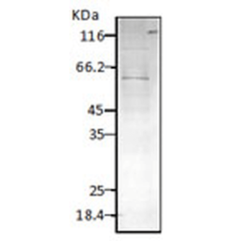
Western Blot analysis of Human Cell line lysates showing detection of Hsp60 protein using Mouse Anti-Hsp60 Monoclonal Antibody, Clone LK-1. Load: 15 μg. Block: 1.5% BSA for 30 minutes at RT. Primary Antibody: Mouse Anti-Hsp60 Monoclonal Antibody at 1:1000 for 2 hours at RT. Secondary Antibody: Sheep Anti-Mouse IgG: HRP for 1 hour at RT.
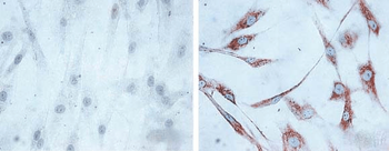
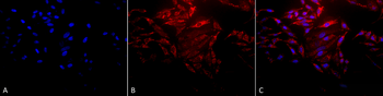
Immunocytochemistry/Immunofluorescence analysis using Mouse Anti-Hsp60 Monoclonal Antibody, Clone LK1, . Tissue: skin Fibroblasts. Species: Human. Fixation: Cold 100% methanol for 30 minutes at -20°C. Primary Antibody: Mouse Anti-Hsp60 Monoclonal Antibody at 1:1000 for 1 hour at RT. Secondary Antibody: DAKO LSAB2 streptavidin-peroxidase system. Counterstain: Mayer Hematoxylin (purple/blue) nuclear stain. Left: control; Right: 24 hours after 7th passage of senescence.
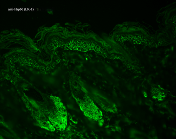
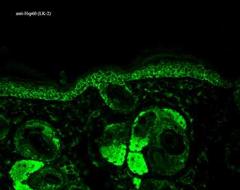
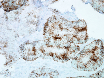
Immunohistochemistry analysis using Mouse Anti-Hsp60 Monoclonal Antibody, Clone LK-1. Tissue: backskin. Species: Mouse. Fixation: Bouin's Fixative and paraffin-embedded. Primary Antibody: Mouse Anti-Hsp60 Monoclonal Antibody at 1:100 for 1 hour at RT. Secondary Antibody: FITC Goat Anti-Mouse (green) at 1:50 for 1 hour at RT. Localization: Epidermis.
- Item 1 of 4
HSP60 Antibody: Biotin [orb151275]
ELISA, ICC, IF, IHC, WB
Bovine, Canine, Gallus, Hamster, Human, Mouse, Rabbit, Rat
Rabbit
Polyclonal
Biotin
100 μg - Item 1 of 4
HSP60 Antibody: Biotin [orb146871]
ELISA, ICC, IF, IHC, WB
Bacteria, Bovine, Canine, E. coli, Fish, Gallus, Guinea pig, H. pylori, Hamster, Human, Insect, Monkey, Mouse, Other, Plant, Porcine, Rabbit, Rat, Yeast
Mouse
Monoclonal
Biotin
200 μg - Item 1 of 2
HSP60 (P. falciparum) Antibody: Biotin [orb152049]
ELISA, ICC, IF, IHC, WB
Bacteria, E. coli
Rabbit
Polyclonal
Biotin
100 μg - Item 1 of 2
Cpn10 Antibody: Biotin [orb152100]
ELISA, ICC, IF, IHC, WB
Bovine, Canine, Frog, Guinea pig, Human, Mouse, Porcine, Rabbit, Rat, Sheep
Rabbit
Polyclonal
Biotin
100 μg