You have no items in your shopping cart.
Cart summary
Item 1 of 3
Item 1 of 3
HSP47 Antibody: Biotin
Catalog Number: orb148127

| Catalog Number | orb148127 |
|---|---|
| Category | Antibodies |
| Description | Mouse monoclonal to Hsp47 (Biotin). Hsp47 is a chaperone protein, member of the superfamily of serine proteinase inhibitors. Also known as SERPINH1, a serine proteinase inhibitor. It is a stress protein that resides in the endoplasmic reticulum, has an active role on the intracellular process of folding, assembly and secretion of pro-collagens. Recent studies have shown the association of on an increased expression of Hsp47 around fibrotic lesions. The identification of a novel biomarker on cell therapies aimed to reduce the progression of fibrotic diseases, could be used potentially as a universal marker, since Hsp47 binds a single substrate. Type I collagen is fundamental during the healing process after a myocardial infarction. It is critical in the position of collagen-produced cells and the assembly of collagen fibrils.. |
| Species/Host | Mouse |
| Clonality | Monoclonal |
| Clone Number | 1C4-1A6 |
| Tested applications | ELISA, ICC, IF, IHC, WB |
| Reactivity | Human |
| Isotype | IgG1 Kappa |
| Immunogen | Human HSP47, full length |
| Concentration | 1 mg/ml |
| Dilution range | WB (1:1000), ICC/IF (1:100) |
| Conjugation | Biotin |
| MW | 47kDa |
| Target | HSP47 |
| Entrez | 871 |
| UniProt ID | P50454 |
| NCBI | NP_001193943 |
| Storage | Conjugated antibodies should be stored according to the product label |
| Buffer/Preservatives | 136.36mM Ethanolamine, and 9.55mM Sodium Bicarbonate in 95.45% PBS |
| Alternative names | SerpinH1 antibody, Colligin antibody, Gp46 antibod Read more... |
| Note | For research use only |
| Application notes | 1 µg/ml of SMC-203 was sufficient for detection of HSP47 in 20 µg of heat shocked HeLa cell lysate by colorimetric immunoblot analysis using Goat anti-mouse IgG:HRP as the secondary antibody. |
| Expiration Date | 12 months from date of receipt. |

Immunocytochemistry/Immunofluorescence analysis using Mouse Anti-Hsp47 Monoclonal Antibody, Clone 1C4-1A6. Tissue: Heat Shocked cervical cancer cells (HeLa). Species: Human. Fixation: 2% Formaldehyde for 20 min at RT. Primary Antibody: Mouse Anti-Hsp47 Monoclonal Antibody at 1:100 for 12 hours at 4°C. Secondary Antibody: FITC Goat Anti-Mouse (green) at 1:200 for 2 hours at RT. Counterstain: DAPI (blue) nuclear stain at 1:40000 for 2 hours at RT. Localization: Endoplasmic reticulum lumen. Cytoplasm. Magnification: 100x. (A) DAPI (blue) nuclear stain. (B) Anti-Hsp47 Antibody. (C) Composite. Heat Shocked at 42°C for 1h.
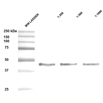
Western Blot analysis of Human Epithelial cell (A431) lysates showing detection of ~47 kDa Hsp47 protein using Mouse Anti-Hsp47 Monoclonal Antibody, Clone 1C4-1A6. Lane 1: MW ladder. Lane 2: Anti-Hsp47 (1:250). Lane 3: Anti-Hsp47 (1:500). Lane 4: Anti-Hsp47 (1:1000). Load: 20 μg. Block: 5% milk + TBST for 1 hour at RT. Primary Antibody: Mouse Anti-Hsp47 Monoclonal Antibody at 1:250 - 1:1000 for 1 hour at RT. Secondary Antibody: HRP Goat Anti-Mouse at 1:50 for 1 hour at RT. Color Development: TMB solution for 10 min at RT. Predicted/Observed Size: ~47 kDa.
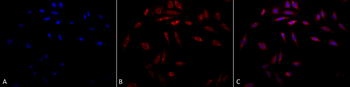
Immunocytochemistry/Immunofluorescence analysis using Mouse Anti-Hsp47 Monoclonal Antibody, Clone 1C4-1A6. Tissue: Heat Shocked cervical cancer cells (HeLa). Species: Human. Fixation: 2% Formaldehyde for 20 min at RT. Primary Antibody: Mouse Anti-Hsp47 Monoclonal Antibody at 1:100 for 12 hours at 4°C. Secondary Antibody: APC Goat Anti-Mouse (red) at 1:200 for 2 hours at RT. Counterstain: DAPI (blue) nuclear stain at 1:40000 for 2 hours at RT. Localization: Endoplasmic reticulum lumen. Cytoplasm. Magnification: 20x. (A) DAPI (blue) nuclear stain. (B) Anti-Hsp47 Antibody. (C) Composite. Heat Shocked at 42°C for 1h.

SERPINH1 Rabbit Polyclonal Antibody (Biotin) [orb2081224]
IP, WB
Canine, Equine, Human, Mouse, Porcine, Rat
Rabbit
Polyclonal
Biotin
100 μl
