You have no items in your shopping cart.
Cart summary
Item 1 of 4
Item 1 of 4
HSP27 Antibody: RPE
Catalog Number: orb151297

| Catalog Number | orb151297 |
|---|---|
| Category | Antibodies |
| Description | Rabbit polyclonal to Hsp27 (RPE). Hsp27s belong to an abundant and ubiquitous family of small heat shock proteins (sHSP). It is an important HSP found in both normal human cells and cancer cells. The basic structure of most sHsps is a homologous and highly conserved amino acid sequence, with an alpha-crystallin domain at the C-terminus and the WD/EPF domain at the less conserved N-terminus. This N-terminus is essential for the development of high molecular oligomers. Hsp27-oligomers consist of stable dimers formed by as many as 8-40 Hsp27 protein monomers. The oligomerization status is connected with the chaperone activity: aggregates of large oligomers have high chaperone activity, whereas dimers have no chaperone activity. HSP27 is localized to the cytoplasm of unstressed cells but can redistribute to the nucleus in response to stress, where it may function to stabilize DNA and/or the nuclear membrane. Other functions include chaperone activity (as mentioned above), thermo tolerance in vivo, inhibition of apoptosis, and signal transduction. Specifically, in vitro, it acts as an ATP-independent chaperone by inhibiting protein aggregation and by stabilizing partially denatured proteins, which ensures refolding of the HSP70 complex. Hsp27 is also involved in the apoptotic signaling pathway because it interferes with the activation of cytochrome c/Apaf-1/dATP complex, thereby inhibiting the activation of procaspase-9. It is also hypothesized that hsp27 may serve some role in cross-bridge formation between actin and myosin. And finally, Hsp27 is also thought to be involved in the process of cell differentiation. The up-regulation of Hsp27 correlates with the rate of phosphorylation and with an increase of large oligomers. It is possible that Hsp27 may play a crucial role in termination of growth.... |
| Species/Host | Rabbit |
| Clonality | Polyclonal |
| Tested applications | ICC, IF, IHC, WB |
| Reactivity | Canine, Fish, Human, Mouse |
| Immunogen | Human HSP27, His tagged |
| Concentration | 1.38 mg/ml |
| Dilution range | WB (1:2000), ICC/IF (1:250) |
| Conjugation | RPE |
| MW | 27kDa |
| Target | HSP27 |
| Entrez | 3315 |
| UniProt ID | P04792 |
| NCBI | NP_001531.1 |
| Storage | Conjugated antibodies should be stored according to the product label |
| Buffer/Preservatives | 95.64mM Phosphate, 2.48mM MES and 2mM EDTA |
| Alternative names | 28kDa heat shock protein antibody, CMT2F antibody, Read more... |
| Note | For research use only |
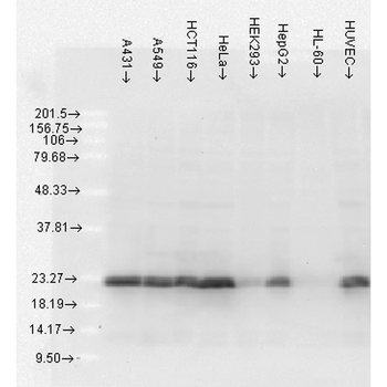
| Application notes | A 1:2000 dilution of SPC-106 was sufficient for detection of HSP27 in 20 µg of HeLa cell lysate by ECL immunoblot analysis. |
| Expiration Date | 12 months from date of receipt. |

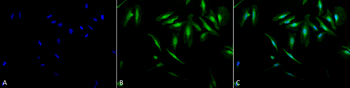
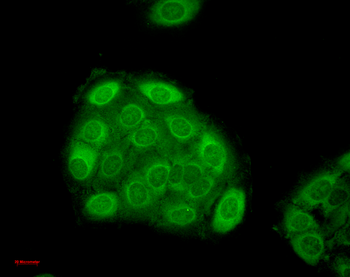
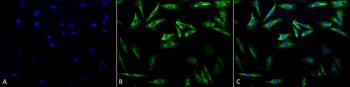
Immunocytochemistry/Immunofluorescence analysis using Rabbit Anti-Hsp27 Polyclonal Antibody. Tissue: Heat Shocked Cervical cancer cell line (HeLa). Species: Human. Fixation: 2% Formaldehyde for 20 min at RT. Primary Antibody: Rabbit Anti-Hsp27 Polyclonal Antibody at 1:250 for 12 hours at 4°C. Secondary Antibody: FITC Goat Anti-Rabbit (green) at 1:200 for 2 hours at RT. Counterstain: DAPI (blue) nuclear stain at 1:40000 for 2 hours at RT. Localization: Cytoplasm. Mitochondrion matrix. Magnification: 100x. (A) DAPI (blue) nuclear stain. (B) Anti-Hsp27 Antibody. (C) Composite. Heat Shocked at 42°C for 1h.
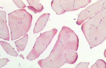
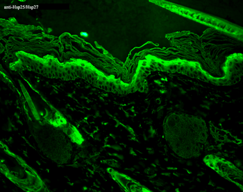
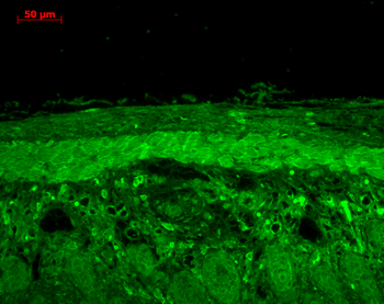
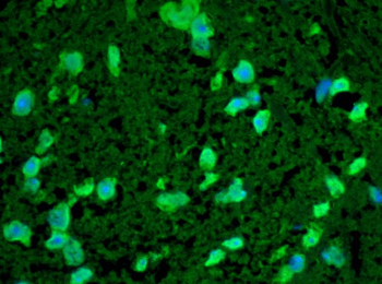
Immunohistochemistry analysis using Rabbit Anti-HSP27 Polyclonal Antibody. Tissue: Skeletal Muscle. Species: Human. Fixation: Formalin fixed paraffin-embedded. Primary Antibody: Rabbit Anti-HSP27 Polyclonal Antibody.
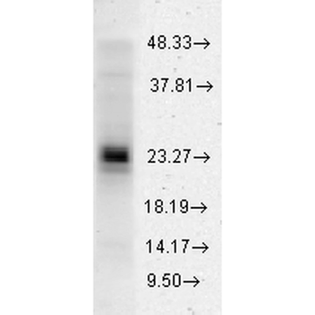
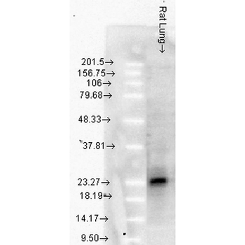
Western blot analysis of Human Cervical cancer cell line (HeLa) lysate showing detection of HSP27 protein using Rabbit Anti-HSP27 Polyclonal Antibody. Load: 15 μgprotein. Block: 1.5% BSA. Primary Antibody: Rabbit Anti-HSP27 Polyclonal Antibody at 1:2000 for 2 hours at RT. Secondary Antibody: Donkey Anti-Rabbit IgG: HRP for 1 hour at RT.
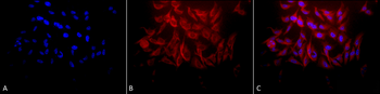
Immunocytochemistry/Immunofluorescence analysis using Rabbit Anti-Hsp27 Polyclonal Antibody. Tissue: Heat Shocked Cervical cancer cell line (HeLa). Species: Human. Fixation: 2% Formaldehyde for 20 min at RT. Primary Antibody: Rabbit Anti-Hsp27 Polyclonal Antibody at 1:250 for 12 hours at 4°C. Secondary Antibody: APC Goat Anti-Rabbit (red) at 1:200 for 2 hours at RT. Counterstain: DAPI (blue) nuclear stain at 1:40000 for 2 hours at RT. Localization: Cytoplasm. Mitochondrion matrix. Magnification: 20x. (A) DAPI (blue) nuclear stain. (B) Anti-Hsp27 Antibody. (C) Composite. Heat Shocked at 42°C for 1h.
- Item 1 of 5
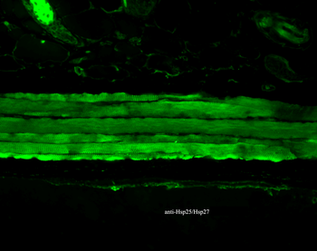
HSP25/HSP27 Antibody: RPE [orb181813]
ICC, IF, IHC, WB
Bovine, Canine, Guinea pig, Hamster, Human, Mouse, Rat, Sheep
Mouse
Monoclonal
RPE
100 μg - Item 1 of 4
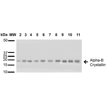
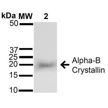
Alpha B Crystallin Antibody: RPE [orb151552]
ICC, IF, IHC, WB
Bovine, Gallus, Human, Mouse, Rat
Rabbit
Polyclonal
RPE
200 μl - Item 1 of 4
- Item 1 of 4
- Item 1 of 2
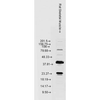
Alpha A Crystallin Antibody: RPE [orb147267]
ICC, IF, IHC, WB
Bovine, Human, Mouse, Rat
Mouse
Monoclonal
RPE
200 μg