You have no items in your shopping cart.
Cart summary
Item 1 of 4
Item 1 of 4
HSC70 (HSP73) Antibody: RPE
Catalog Number: orb147352

| Catalog Number | orb147352 |
|---|---|
| Category | Antibodies |
| Description | Mouse monoclonal to Hsc70 (RPE). Hsp70 genes encode abundant heat-inducible 70-kDa hsps (hsp70s). In most eukaryotes hsp70 genes exist as part of a multigene family. They are found in most cellular compartments of eukaryotes including nuclei, mitochondria, chloroplasts, the endoplasmic reticulum and the cytosol, as well as in bacteria. The genes show a high degree of conservation, having at least 5O% identity. The N-terminal two thirds of hsp70s are more conserved than the C-terminal third. Hsp70 binds ATP with high affinity and possesses a weak ATPase activity which can be stimulated by binding to unfolded proteins and synthetic peptides. When hsc70 (constitutively expressed) present in mammalian cells was truncated, ATP binding activity was found to reside in an N-terminal fragment of 44 kDa which lacked peptide binding capacity. Polypeptide binding ability therefore resided within the C-terminal half. The structure of this ATP binding domain displays multiple features of nucleotide binding proteins. When cells are subjected to metabolic stress (e. g. , heat shock) a member of the hsp 70 family, hsp 70 (hsp72), is expressed; hsp 70 is highly related to hsc70 (>90% sequence identity). Constitutively expressed hsc70 rapidly forms a stable complex with the highly inducible hsp70 in cells following heat shock. The interaction of hsc70 with hsp 70 is regulated by ATP. These two heat shock proteins move together in the cell experiencing stress. Furthermore, research on hsc70 has implicates it with a role in facilitating the recovery of centrosomal structure and function after heat shock.... |
| Species/Host | Mouse |
| Clonality | Monoclonal |
| Clone Number | 1F2-H5 |
| Tested applications | ICC, IF, IHC, WB |
| Reactivity | Human, Mouse, Rat |
| Isotype | IgG2a Kappa |
| Immunogen | Full length human HSC70 |
| Concentration | 1 mg/ml |
| Dilution range | WB (1:1000), ICC/IF (1:100) |
| Conjugation | RPE |
| MW | 73kDa |
| Target | HSC70 (HSP73) |
| Entrez | 3312 |
| UniProt ID | P11142 |
| NCBI | NP_006588.1 |
| Storage | Conjugated antibodies should be stored according to the product label |
| Buffer/Preservatives | 95.64mM Phosphate, 2.48mM MES and 2mM EDTA |
| Alternative names | HSC54 antibody, HSC71 antibody, HSC73 antibody, HS Read more... |
| Note | For research use only |
| Application notes | 1 µg/ml of SMC-151 was sufficient for detection of HSC70 in 10 µg of HeLa lysate by colorimetric immunoblot analysis using Goat anti-mouse IgG:HRP as the secondary antibody. |
| Expiration Date | 12 months from date of receipt. |

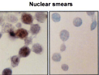
Immunocytochemistry/Immunofluorescence analysis using Mouse Anti-Hsc70 (Hsp73) Monoclonal Antibody, Clone 1F2-H5. Tissue: Heat Shocked cervical cancer cells (HeLa). Species: Human. Fixation: 2% Formaldehyde for 20 min at RT. Primary Antibody: Mouse Anti-Hsc70 (Hsp73) Monoclonal Antibody at 1:100 for 12 hours at 4°C. Secondary Antibody: FITC Goat Anti-Mouse (green) at 1:200 for 2 hours at RT. Counterstain: DAPI (blue) nuclear stain at 1:40000 for 2 hours at RT. Localization: Cytoplasm. Melanosome. Localizes to nucleus upon heat shock. Magnification: 100x. (A) DAPI (blue) nuclear stain. (B) Anti-Hsc70 (Hsp73) Antibody. (C) Composite. Heat Shocked at 42°C for 1h.
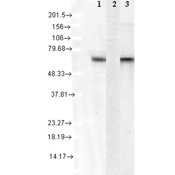
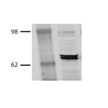
Western Blot analysis of Human Cell lysates showing detection of Hsc70 protein using Mouse Anti-Hsc70 Monoclonal Antibody, Clone 1F2-H5. Load: 15 μg. Block: 1.5% BSA for 30 minutes at RT. Primary Antibody: Mouse Anti-Hsc70 Monoclonal Antibody at 1:1000 for 2 hours at RT. Secondary Antibody: Sheep Anti-Mouse IgG: HRP for 1 hour at RT. 1: mix of 10 different human cell lines, 2: Hsp72 recombinant protein, and 3: Hsc70 (Hsp73) recombinant protein.

Immunocytochemistry/Immunofluorescence analysis using Mouse Anti-Hsc70 (Hsp73) Monoclonal Antibody, Clone 1F2-H5. Tissue: Heat Shocked cervical cancer cells (HeLa). Species: Human. Fixation: 2% Formaldehyde for 20 min at RT. Primary Antibody: Mouse Anti-Hsc70 (Hsp73) Monoclonal Antibody at 1:100 for 12 hours at 4°C. Secondary Antibody: R-PE Goat Anti-Mouse (yellow) at 1:200 for 2 hours at RT. Counterstain: DAPI (blue) nuclear stain at 1:40000 for 2 hours at RT. Localization: Cytoplasm. Melanosome. Localizes to nucleus upon heat shock. Magnification: 20x. (A) DAPI (blue) nuclear stain. (B) Anti-Hsc70 (Hsp73) Antibody. (C) Composite. Heat Shocked at 42°C for 1h.
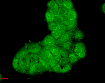
Immunocytochemistry/Immunofluorescence analysis using Mouse Anti-Hsc70 Monoclonal Antibody, Clone 1F2-H5. Tissue: HaCaT cells. Species: Human. Fixation: Cold 100% methanol for 10 minutes at -20°C. Primary Antibody: Mouse Anti-Hsc70 Monoclonal Antibody at 1:100 for 1 hour at RT. Secondary Antibody: FITC Goat Anti-Mouse (green) at 1:50 for 1 hour at RT. Localization: Bright cytoplasmic staining, duller nuclear staining.
- Item 1 of 6
HSP70/HSC70 Antibody: RPE [orb146791]
ICC, IF, IHC, WB
Bovine, Canine, Drosophila, Fish, Frog, Gallus, Guinea pig, Hamster, Human, Mouse, Other, Porcine, Rabbit, Rat, Sheep, Yeast
Mouse
Monoclonal
RPE
200 μg - Item 1 of 3
HSP70 Antibody: RPE [orb147573]
ICC, IF, IHC, WB
Amphibian, Bacteria, Crustacean, Drosophila, Fish, Gallus, Human, Mouse, Rat, Saccharomyces, Yeast
Mouse
Monoclonal
RPE
100 μg