You have no items in your shopping cart.
Cart summary
Item 1 of 4
Item 1 of 4
HRAS Antibody (Center)
Catalog Number: orb1928912

| Catalog Number | orb1928912 |
|---|---|
| Category | Antibodies |
| Description | Purified Rabbit Polyclonal Antibody (Pab) |
| Species/Host | Rabbit |
| Clonality | Polyclonal |
| Clone Number | RB15622 |
| Tested applications | IF, IHC-P, WB |
| Predicted Reactivity | Mouse, Rat |
| Reactivity | Human |
| Isotype | Rabbit IgG |
| Dilution range | IF: 1:10~50, WB: 1:1000, WB: 1:500, IHC-P: 1:10~50 |
| Form/Appearance | Purified polyclonal antibody supplied in PBS with 0.09% (W/V) sodium azide. This antibody is prepared by Saturated Ammonium Sulfate (SAS) precipitation followed by dialysis against PBS. |
| Conjugation | Unconjugated |
| MW | 21298 Da |
| Target | This HRAS antibody is generated from rabbits immunized with a KLH conjugated synthetic peptide between 104-128 amino acids from the Central region of human HRAS. |
| UniProt ID | P01112 |
| NCBI | NP_001123914.1, NP_789765.1, NP_005334.1 |
| Storage | Maintain refrigerated at 2-8°C for up to 2 weeks. For long term storage store at -20°C in small aliquots to prevent freeze-thaw cycles |
| Alternative names | GTPase HRas, H-Ras-1, Ha-Ras, Transforming protein Read more... |
| Note | For research use only |
| Expiration Date | 12 months from date of receipt. |

Western blot analysis of anti-HRAS Antibody (Center) in Jurkat cell line lysates (35 ug/lane). HRAS (arrow) was detected using the purified Pab.
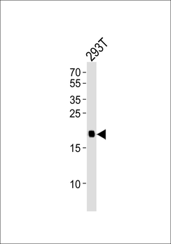
Western blot analysis of lysate from 293T cell line, using HRAS Antibody (Center). Diluted at 1:1000. A goat anti-rabbit IgG H&L (HRP) at 1:10000 dilution was used as the secondary antibody. Lysate at 20 ug.
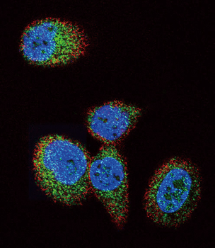
Confocal immunofluorescent analysis of HRAS Antibody (Center) with MCF-7 cell followed by Alexa Fluor 488-conjugated goat anti-rabbit lgG (green).Actin filaments have been labeled with Alexa Fluor 555 phalloidin (red).DAPI was used to stain the cell nuclear (blue).
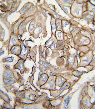
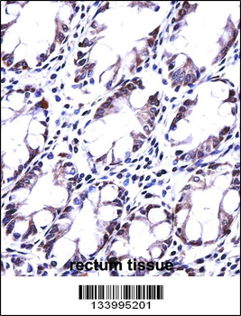
Formalin-fixed and paraffin-embedded human lung carcinoma tissue reacted with HRAS antibody (Center), which was peroxidase-conjugated to the secondary antibody, followed by DAB staining. This data demonstrates the use of this antibody for immunohistochemistry; clinical relevance has not been evaluated.
- Item 1 of 2
- Item 1 of 1