You have no items in your shopping cart.
Cart summary
Item 1 of 4
Item 1 of 4
HP Antibody (Center)
Catalog Number: orb1928202

| Catalog Number | orb1928202 |
|---|---|
| Category | Antibodies |
| Description | Affinity Purified Rabbit Polyclonal Antibody (Pab) |
| Species/Host | Rabbit |
| Clonality | Polyclonal |
| Clone Number | RB21463 |
| Tested applications | FC, IHC-P, WB |
| Reactivity | Human, Mouse |
| Isotype | Rabbit IgG |
| Dilution range | WB: 1:1000, WB: 1:8000, IHC-P: 1:25, FC: 1:10~50 |
| Form/Appearance | Purified polyclonal antibody supplied in PBS with 0.09% (W/V) sodium azide. This antibody is purified through a protein A column, followed by peptide affinity purification. |
| Conjugation | Unconjugated |
| MW | 45205 Da |
| Target | This HP antibody is generated from rabbits immunized with a KLH conjugated synthetic peptide between 295-322 amino acids from the Central region of human HP. |
| UniProt ID | P00738 |
| NCBI | NP_005134.1, NP_001119574.1 |
| Storage | Maintain refrigerated at 2-8°C for up to 2 weeks. For long term storage store at -20°C in small aliquots to prevent freeze-thaw cycles |
| Alternative names | Haptoglobin, Zonulin, Haptoglobin alpha chain, Hap Read more... |
| Note | For research use only |
| Expiration Date | 12 months from date of receipt. |

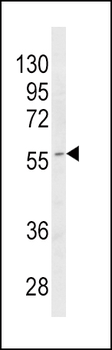
Western blot analysis of HP (arrow) using rabbit polyclonal HP Antibody (Center). 293 cell lysates (2 ug/lane) either nontransfected (Lane 1) or transiently transfected with the HP gene (Lane 2).
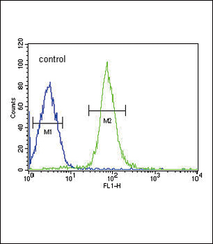
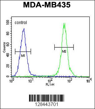
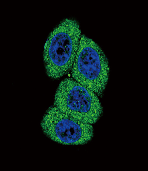
HP Antibody (Center) flow cytometric analysis of HepG2 cells (right histogram) compared to a negative control cell (left histogram). FITC-conjugated goat-anti-rabbit secondary antibodies were used for the analysis.

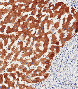
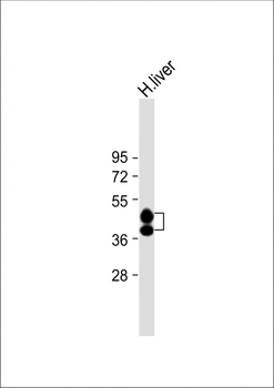
Anti-HP Antibody (Center) at 1:8000 dilution + Human liver lysate. Lysates/proteins at 20 µg per lane. Secondary Goat Anti-Rabbit IgG, (H+L), Peroxidase conjugated at 1/10000 dilution. Predicted band size: 45, 38 kDa. Blocking/Dilution buffer: 5% NFDM/TBST.
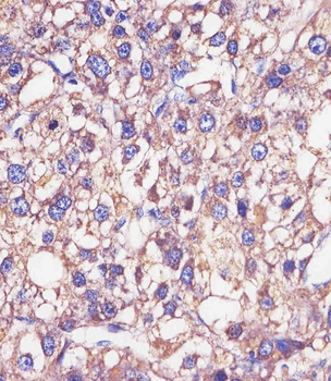
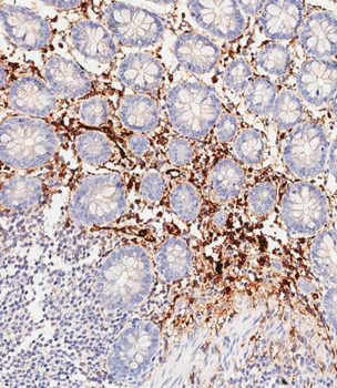
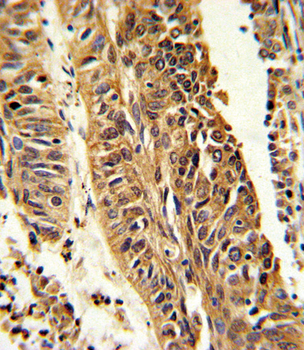
Staining HP in human epatocarcinoma sections by Immunohistochemistry (IHC-P - paraformaldehyde-fixed, paraffin-embedded sections). Tissue was fixed with formaldehyde and blocked with 3% BSA for 0.5 hour at room temperature; antigen retrieval was by heat mediation with a citrate buffer (pH6). Samples were incubated with primary antibody (1/25) for 1 hours at 37°C. A undiluted biotinylated goat polyvalent antibody was used as the secondary antibody.
- Item 1 of 5
- Item 1 of 5
- Item 1 of 4
- Item 1 of 3
- Item 1 of 3