You have no items in your shopping cart.
Cart summary
Item 1 of 5
Item 1 of 5
HO-1 Antibody: APC
Catalog Number: orb147091

| Catalog Number | orb147091 |
|---|---|
| Category | Antibodies |
| Description | Mouse monoclonal to HO-1 (APC). Heme-oxygenase is a ubiquitous enzyme that catalyzes Heme-oxygenase is a ubiquitous enzyme that catalyzes... |
| Species/Host | Mouse |
| Clonality | Monoclonal |
| Clone Number | 1F12-A6 |
| Tested applications | ICC, IF, IHC |
| Reactivity | Bovine, Canine, Guinea pig, Hamster, Human, Monkey, Mouse, Porcine, Rabbit, Rat |
| Isotype | IgG1 Kappa |
| Immunogen | Human HO-1 synthetic peptide, amino acids 1-30 |
| Concentration | 1 mg/ml |
| Dilution range | WB (1:1000), IHC (1:100), ICC/IF (1:100) |
| Conjugation | APC |
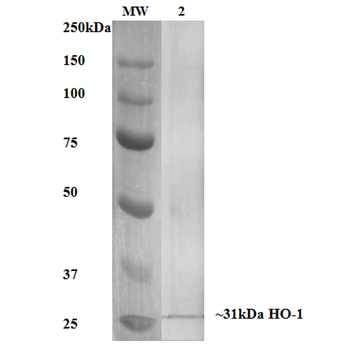
| MW | 32kDa |
| Target | HO-1 |
| Entrez | 3162 |
| UniProt ID | P09601 |
| NCBI | NP_002124.1 |
| Storage | Conjugated antibodies should be stored according to the product label |
| Buffer/Preservatives | 95.64mM Phosphate, 2.48mM MES and 2mM EDTA |
| Alternative names | HSP32 antibody, HMOX1 antibody, Heme oxygenase 1 a Read more... |
| Note | For research use only |
| Application notes | 1 µg/ml was sufficient for detection of HO-1 in 10 µg of mixed human cell line lysate by colorimetric immunoblot analysis using Goat Anti-Mouse IgG:HRP as the secondary. |
| Expiration Date | 12 months from date of receipt. |

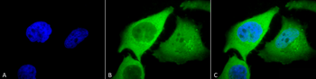
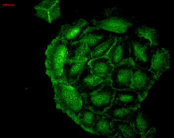
Immunocytochemistry/Immunofluorescence analysis using Mouse Anti-HO-1 Monoclonal Antibody, Clone 1F12-A6. Tissue: Cervical cancer cell line (HeLa). Species: Human. Fixation: 2% Formaldehyde for 20 min at RT. Primary Antibody: Mouse Anti-HO-1 Monoclonal Antibody at 1:100 for 12 hours at 4°C. Secondary Antibody: FITC Goat Anti-Mouse (green) at 1:200 for 2 hours at RT. Counterstain: DAPI (blue) nuclear stain at 1:40000 for 2 hours at RT. Localization: Microsome. Endoplasmic reticulum. Localizes to the nucleus upon hypoxia. Magnification: 100x. (A) DAPI (blue) nuclear stain. (B) Anti-HO-1 Antibody. (C) Composite.
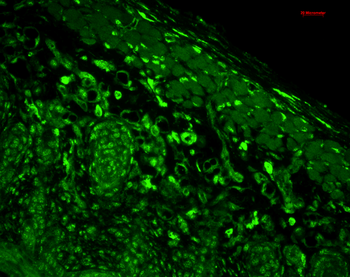
Immunohistochemistry analysis using Mouse Anti-HO-1 Monoclonal Antibody, Clone 1F12-A6. Tissue: backskin. Species: Mouse. Fixation: Bouin's Fixative and paraffin-embedded. Primary Antibody: Mouse Anti-HO-1 Monoclonal Antibody at 1:100 for 1 hour at RT. Secondary Antibody: FITC Goat Anti-Mouse (green) at 1:50 for 1 hour at RT. Localization: muscle, dermis, hair follicles, epidermis: nuclear everywhere and some cytoplasmic staining.
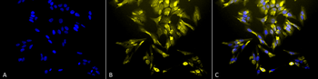
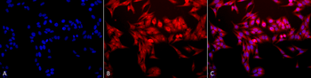
Immunocytochemistry/Immunofluorescence analysis using Mouse Anti-HO-1 Monoclonal Antibody, Clone 1F12-A6. Tissue: Cervical cancer cell line (HeLa). Species: Human. Fixation: 2% Formaldehyde for 20 min at RT. Primary Antibody: Mouse Anti-HO-1 Monoclonal Antibody at 1:100 for 12 hours at 4°C. Secondary Antibody: R-PE Goat Anti-Mouse (yellow) at 1:200 for 2 hours at RT. Counterstain: DAPI (blue) nuclear stain at 1:40000 for 2 hours at RT. Localization: Microsome. Endoplasmic reticulum. Localizes to the nucleus upon hypoxia. Magnification: 20x. (A) DAPI (blue) nuclear stain. (B) Anti-HO-1 Antibody. (C) Composite.
Immunocytochemistry/Immunofluorescence analysis using Mouse Anti-HO-1 Monoclonal Antibody, Clone 1F12-A6. Tissue: HaCaT cells. Species: Human. Fixation: Cold 100% methanol for 10 minutes at -20°C. Primary Antibody: Mouse Anti-HO-1 Monoclonal Antibody at 1:100 for 1 hour at RT. Secondary Antibody: FITC Goat Anti-Mouse (green) at 1:50 for 1 hour at RT. Localization: Cell-cell border staining in epidermis, punctuate nuclear staining.
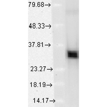
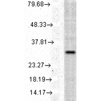
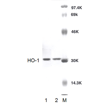
Western Blot analysis of Human Cervical cancer cell line (HeLa) lysate showing detection of HO-1 protein using Mouse Anti-HO-1 Monoclonal Antibody, Clone 1F12-A6. Load: 15 μg. Block: 1.5% BSA for 30 minutes at RT. Primary Antibody: Mouse Anti-HO-1 Monoclonal Antibody at 1:1000 for 2 hours at RT. Secondary Antibody: Sheep Anti-Mouse IgG: HRP for 1 hour at RT.
- Item 1 of 4
- Item 1 of 2
- Item 1 of 2

Heme Oxygenase 1 Rabbit Polyclonal Antibody (APC) [orb1006487]
IF
Bovine, Canine, Equine, Gallus, Guinea pig, Rabbit, Sheep
Human, Mouse, Porcine, Rat
Rabbit
Polyclonal
APC
100 μl