You have no items in your shopping cart.
Cart summary
Item 1 of 16
Item 1 of 16

| Catalog Number | orb195321 |
|---|---|
| Category | Antibodies |
| Description | HMGB1 antibody |
| Species/Host | Rabbit |
| Clonality | Polyclonal |
| Tested applications | ELISA, ICC, IF, IHC-P, WB |
| Reactivity | Bovine, Human, Mouse, Rat |
| Isotype | IgG |
| Immunogen | KLH conjugated synthetic peptide derived from human HMGB1. Please contact us for the exact immunogen sequence. The peptide is available as orb374832. |
| Concentration | - 100 μg (in 200 μl): 0.5 mg/ml- 200 μg (in 400 μl): 0.5 mg/ml |
| Dilution range | WB: 1:5000, IF/ICC: 1:1000, IHC-P: 1:1000 |
| Form/Appearance | 10 mM PBS, 0.02% sodium azide |
| Purity | Polyclonal antibodies are purified by peptide affinity chromatography |
| Conjugation | Unconjugated |
| MW | 24 kDa |
| Target | HMGB1 |
| Entrez | 3146 |
| UniProt ID | P63158, P09429, P63159 |
| NCBI | 002119, 11, 41, 002128 |
| Storage | Maintain refrigerated at 2-8°C for up to 2 weeks. For long term storage store at -20°C in small aliquots to prevent freeze-thaw cycles. |
| Alternative names | anti Amphoterin antibody, anti Chromosomal protein Read more... |
| Note | For research use only |
| Expiration Date | 12 months from date of receipt. |
Filter by Applications
Filter by Reactivity
F Li, N Qin, Y Yu, R Dong, X Li, S Gong, Z Zeng, L Huang, H Yang TREM-1 inhibition or ondansetron administration ameliorates NLRP3 inflammasome and pyroptosis in traumatic brain injury-induced acute lung injury Intensive Care Medicine, (2024)
Applications
WB
Reactivity
Rat
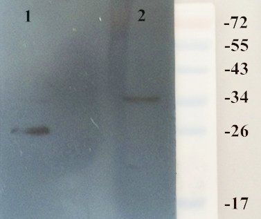
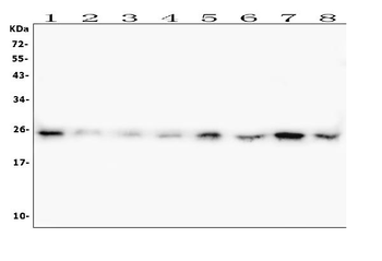
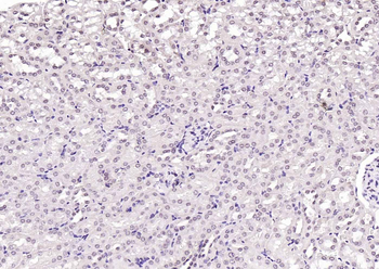
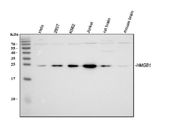
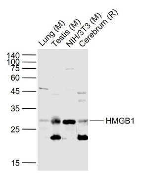
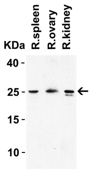
WB analysis of rat kidney (Lane 1), brain (Lane 2) using HMGB1 antibody (Dilution at 1:5000)
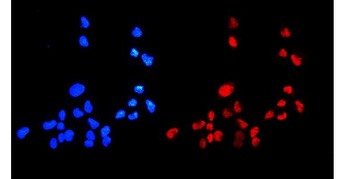
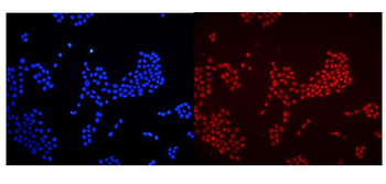
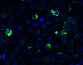
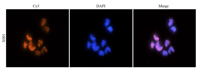
Immunofluorescence analysis of mouse sp20 mouse myeloma cells tissue using anti-HMGB1 (dilution of primary antibody - 1:300)
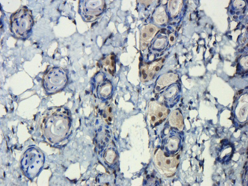
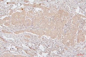
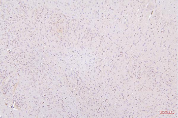
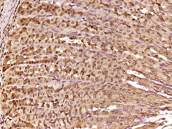
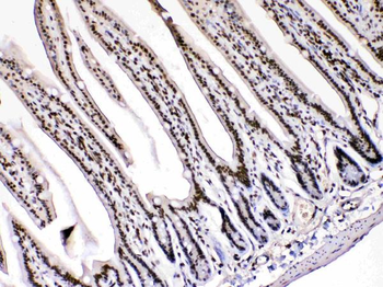
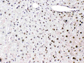
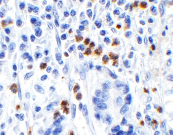
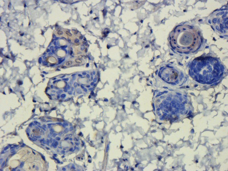
IHC-P staining of mouse skin tissue using HMGB1 antibody (dilution at 1:300)
Immunofluorescence image of mouse sp23 mouse myeloma cells tissue using anti-HMGB1 (dilution at 1:300)
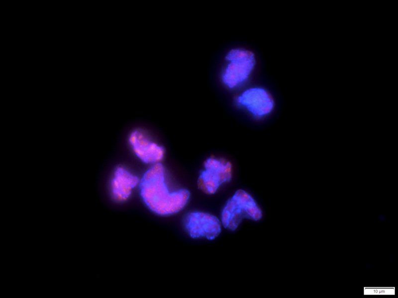
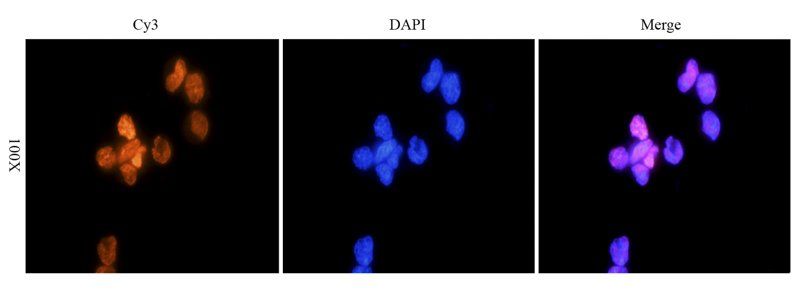
Immunofluorescence image of mouse sp22 mouse myeloma cells tissue using HMGB1 antibody (dilution at 1:1000)
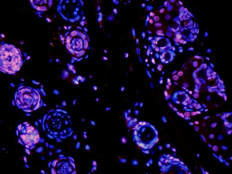
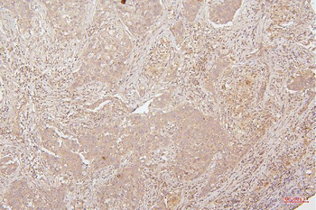
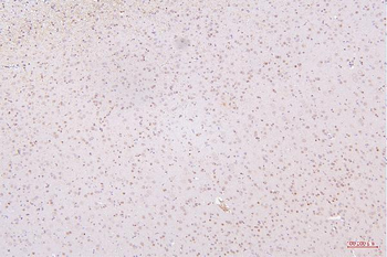
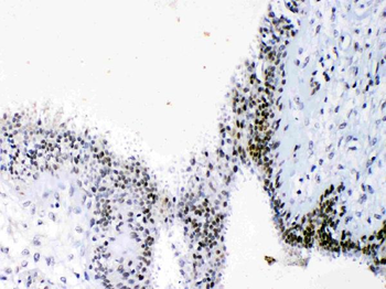
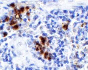
IF image of rat skin tissue using HMGB1 antibody (primary antibody at 1:1000)
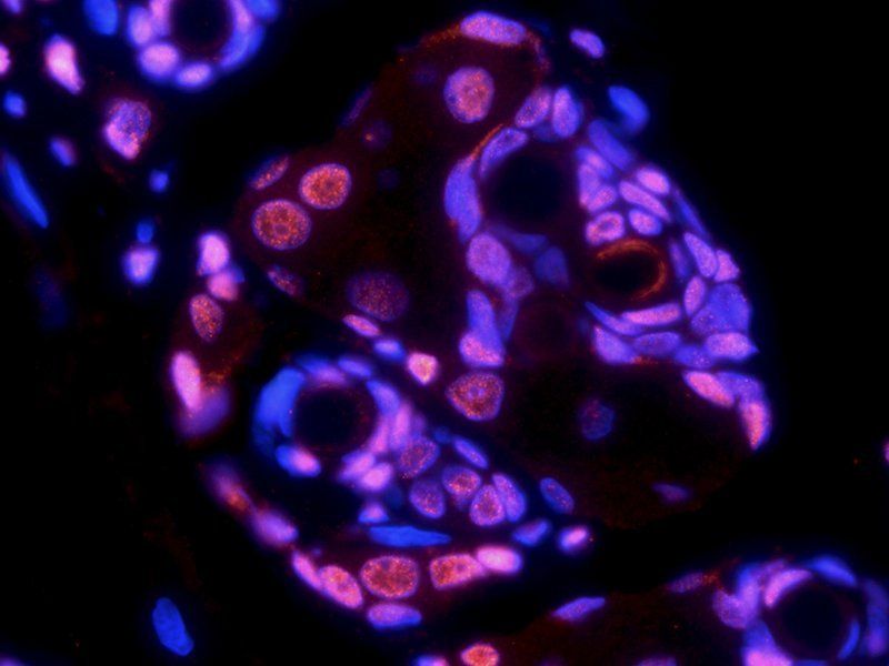
Immunofluorescence image of rat skin tissue using anti-HMGB1 (dilution at 1:1000)

Immunofluorescence image of mouse sp22 mouse myeloma cells tissue using anti-HMGB1 (dilution at 1:300)
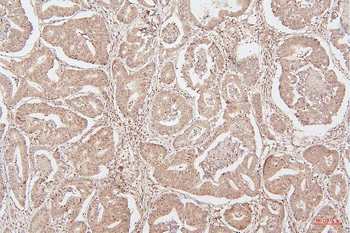
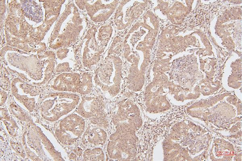
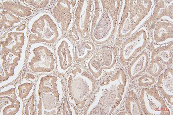
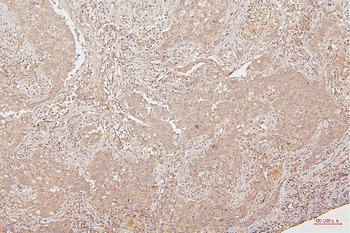
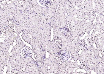
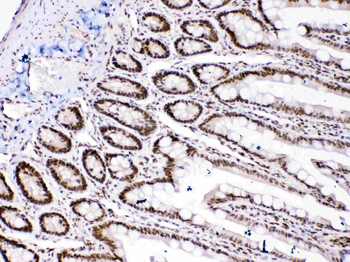
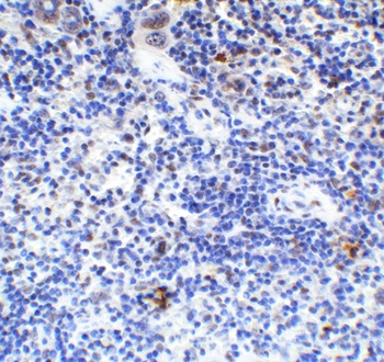
IHC-P staining of rat skin tissue using anti-HMGB1 (dilution at 1:1000)
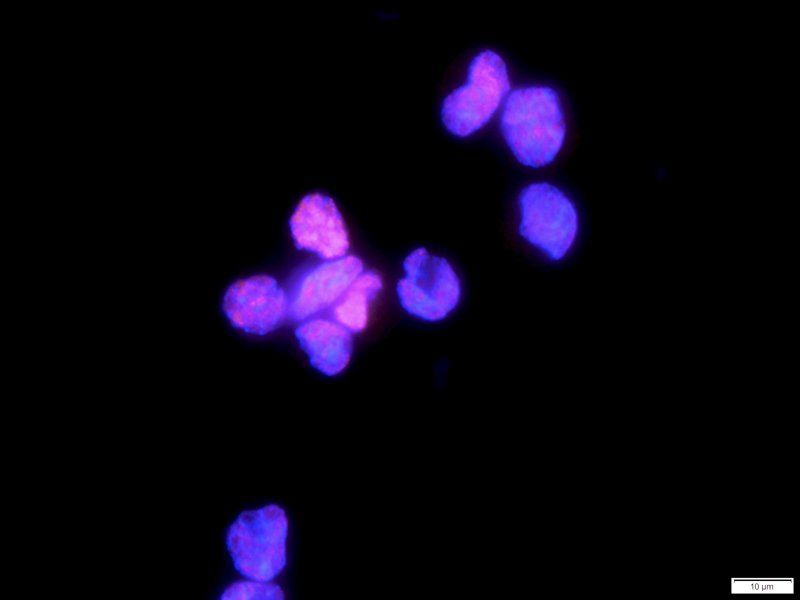
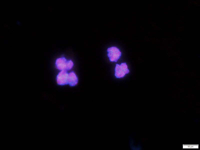
IF analysis of mouse sp21 mouse myeloma cells tissue using HMGB1 antibody (dilution of primary antibody at 1:300)
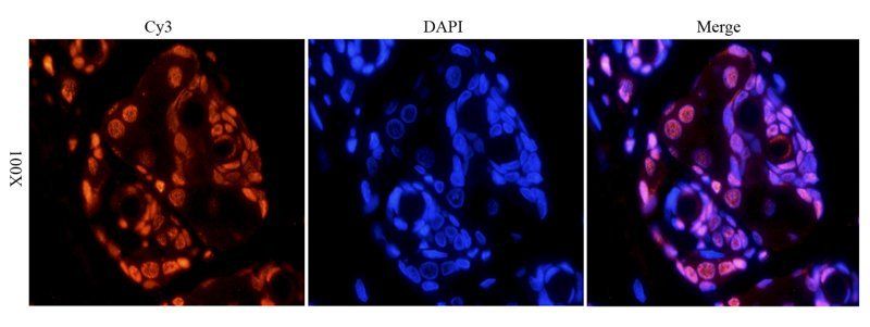
Immunofluorescence analysis of rat skin tissue using HMGB1 antibody (dilution of primary antibody - 1:1000)
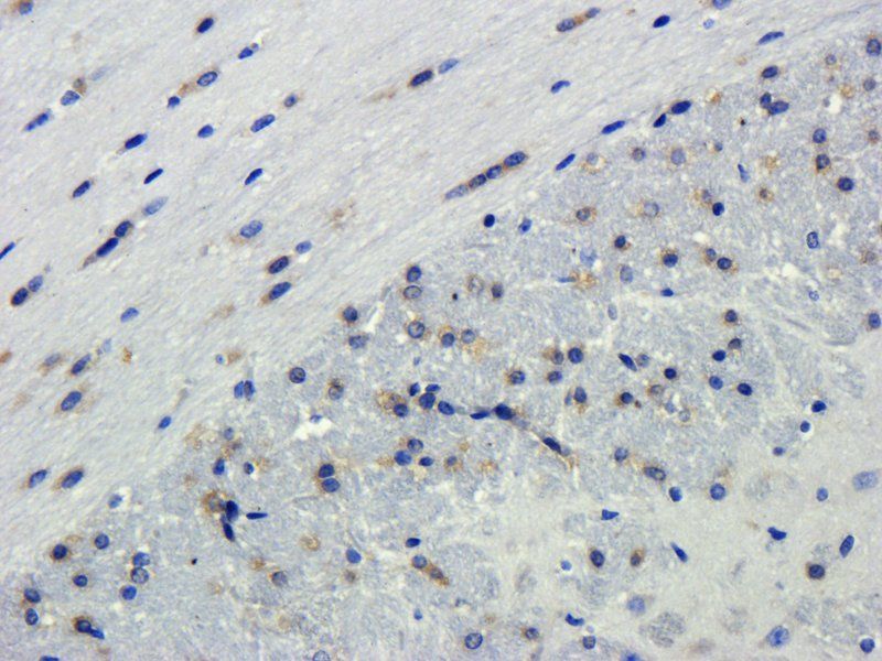
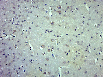
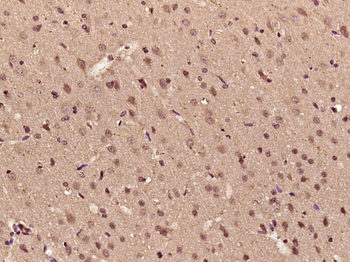
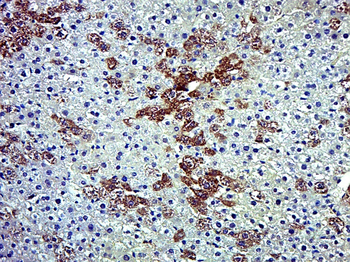
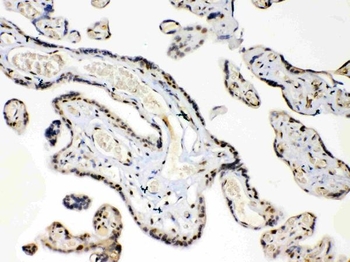
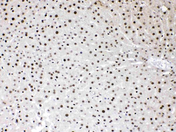
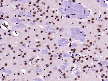
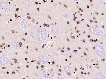
Immunohistochemical staining of paraffin embedded mouse brain tissue using HMGB1 antibody (primary antibody at 1:1000)
Immunofluorescence analysis of mouse sp21 mouse myeloma cells tissue using anti-HMGB1 (dilution of primary antibody - 1:1000)
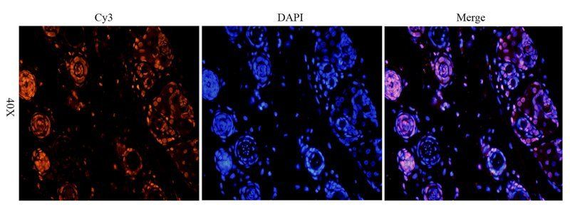
IF image of rat skin tissue using anti-HMGB1 (primary antibody at 1:1000)
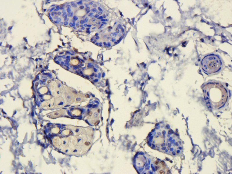
Immunohistochemical staining of rat skin tissue using HMGB1 antibody (dilution of primary antibody - 1:300)
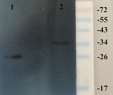
Western blot analysis of rat kidney (Lane 1), rat brain (Lane 2) using HMGB1 antibody (dilution at 1:200)
- Item 1 of 14
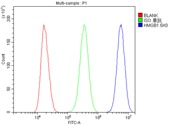
Anti-HMGB1 Antibody (monoclonal, 5H3) [orb570317]
FC, IHC, WB
Human, Monkey, Mouse, Rat
Mouse
Monoclonal
Unconjugated
10 μg, 100 μg - Item 1 of 9
HMGB1 Rabbit Polyclonal Antibody [orb500823]
FC, IF, IHC-Fr, IHC-P
Bovine, Canine, Equine, Porcine
Human, Mouse, Rat
Rabbit
Polyclonal
Unconjugated
100 μl, 50 μl, 200 μl - Item 1 of 11
Anti-HMGB1 Antibody [orb389479]
FC, ICC, IF, IHC, WB
Human, Mouse, Rat
Rabbit
Polyclonal
Unconjugated
10 μg, 100 μg - Item 1 of 9
HMGB1 Rabbit Polyclonal Antibody [orb704527]
ELISA, IF, IHC-Fr, IHC-P, WB
Mouse, Rat
Human, Mouse, Rat
Rabbit
Polyclonal
Unconjugated
100 μl, 50 μl - Item 1 of 8
HMGB1 Antibody [orb1239440]
ELISA, IF, IHC-P, WB
Gallus, Porcine
Human, Mouse, Rat
Rabbit
Polyclonal
Unconjugated
0.1 mg