You have no items in your shopping cart.
HMGB1 Antibody
SKU: orb1239440
Featured
Description
Images & Validation
−Item 1 of 8
| Tested Applications | ELISA, IF, IHC-P, WB |
|---|---|
| Reactivity | Human, Mouse, Rat |
| Predicted Reactivity | Gallus, Porcine |
Key Properties
−| Antibody Type | Primary Antibody |
|---|---|
| Host | Rabbit |
| Clonality | Polyclonal |
| Isotype | IgG |
| Immunogen | Anti-HMGB1 antibody (orb1239440) was raised against a peptide corresponding to 19 amino acids near the center of human HMGB1. The immunogen is located within 70-120 amino acids of HMGB1. |
| Target | HMGB1 |
| Molecular Weight | Predicted: 25 kDa Observed: 25 kDa |
| Purification | HMGB1 antibody is affinity chromatography purified via peptide column. |
| Conjugation | Unconjugated |
Storage & Handling
−| Storage | Antibody can be stored at 4°C up to one year. Antibodies should not be exposed to prolonged high temperatures |
|---|---|
| Form/Appearance | Liquid |
| Buffer/Preservatives | HMGB1 Antibody is supplied in PBS containing 0.02% sodium azide. |
| Concentration | 1 mg/mL |
| Disclaimer | For research use only |
Alternative Names
−HMGB1 Antibody: HMG1, HMG3, SBP-1, HMG1, High mobility group protein B1, High mobility group protein 1, HMG-1
Similar Products
−- Item 1 of 16
HMGB1 antibody [orb195321]
ELISA, ICC, IF, IHC-P, WB
Bovine, Human, Mouse, Rat
Rabbit
Polyclonal
Unconjugated
100 μg - Item 1 of 14
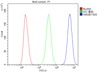
HMGB1 Antibody (monoclonal, 5H3) [orb570317]
FC, IHC, WB
Human, Monkey, Mouse, Rat
Mouse
Monoclonal
Unconjugated
100 μg - Item 1 of 11
- Item 1 of 9
HMGB1 Rabbit Polyclonal Antibody [orb500823]
FC, ICC, IF, IHC-Fr, IHC-P
Bovine, Canine, Equine, Porcine
Human, Mouse, Rat
Rabbit
Polyclonal
Unconjugated
50 μl, 100 μl, 200 μl - Item 1 of 7
HMGB1 Rabbit Polyclonal Antibody [orb704161]
IF, IHC-Fr, IHC-P
Bovine, Canine, Equine, Porcine
Human, Mouse, Rat
Rabbit
Polyclonal
Unconjugated
200 μl, 100 μl, 50 μl


Quality Guarantee
Explore bioreagents carefree to elevate your research. All our products are rigorously tested for performance. If a product does not perform as described on its datasheet, our scientific support team will provide expert troubleshooting, a prompt replacement, or a refund. For full details, please see our Terms & Conditions and Buying Guide. Contact us at [email protected].

KO Validation in HeLa Cells. Loading: 10 µg of HeLa WT cell lysates or HMGB1 KO cell lysates. Antibodies: HMGB1 (0.5 µg/mL) and beta-actin orb1240312 (1 µg/mL), 1 h incubation at RT in 5% NFDM/TBST. Secondary: Goat Anti-Rabbit IgG HRP conjugate at 1:10000 dilution.
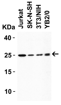
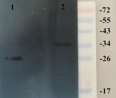
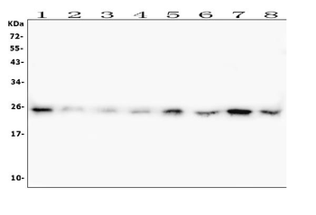
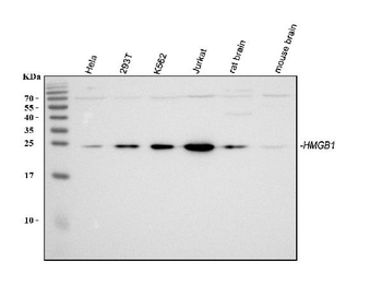
WB Validation in Human, Mouse and Rat Cell Lines. Loading: 15 µg of lysate Antibodies: HMGB1 orb1239440, 1 µg/mL, 1 h incubation at RT in 5% NFDM/TBST. Secondary: Goat Anti-Rabbit IgG HRP conjugate at 1:10000 dilution.
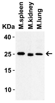
WB Validation in Mouse Tissues. Loading: 15 µg of lysate Antibodies: HMGB1 orb1239440, 1 µ g/mL, 1 h incubation at RT in 5% NFDM/TBST. Secondary: Goat Anti-Rabbit IgG HRP conjugate at 1:10000 dilution.
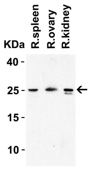
WB Validation in Rat Tissues. Loading: 15 µg of lysate Antibodies: HMGB1 orb1239440, 1 µg/mL, 1 h incubation at RT in 5% NFDM/TBST. Secondary: Goat Anti-Rabbit IgG HRP conjugate at 1:10000 dilution.
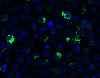
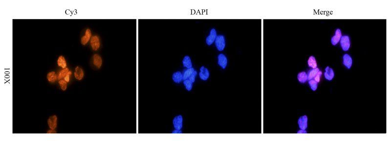
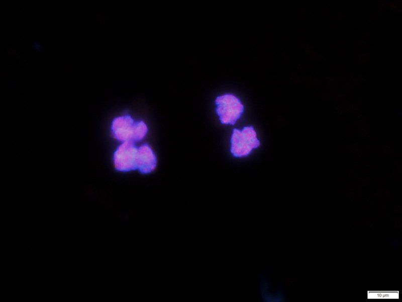
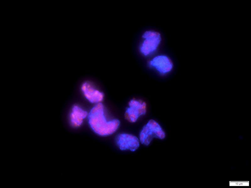
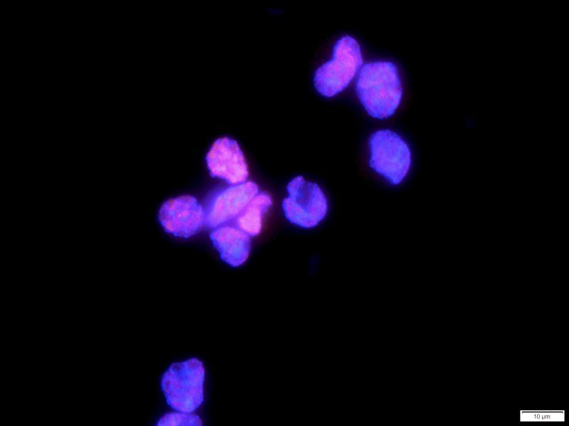
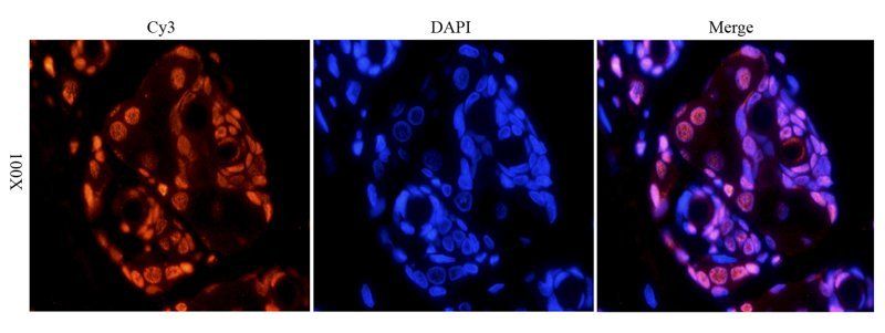
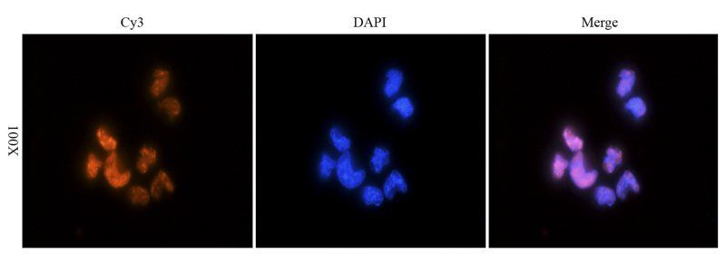
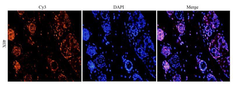
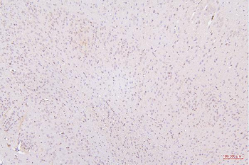
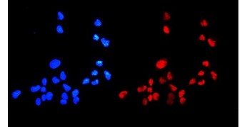
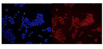
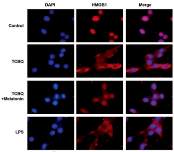
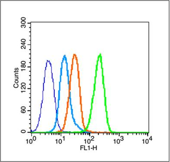
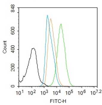
Immunofluorescence Validation of HMGB1 in HL60 Cells. Immunofluorescent analysis of 4% paraformaldehyde-fixed HL60 cells labeling HMGB1 with orb1239440 at 20 µg /mL, followed by goat anti-rabbit IgG secondary antibody at 1/500 dilution (green) and DAPI antibody (blue).
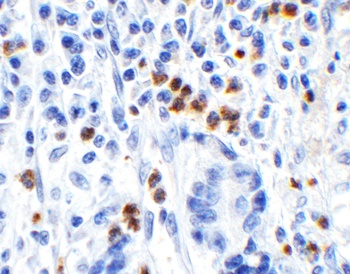
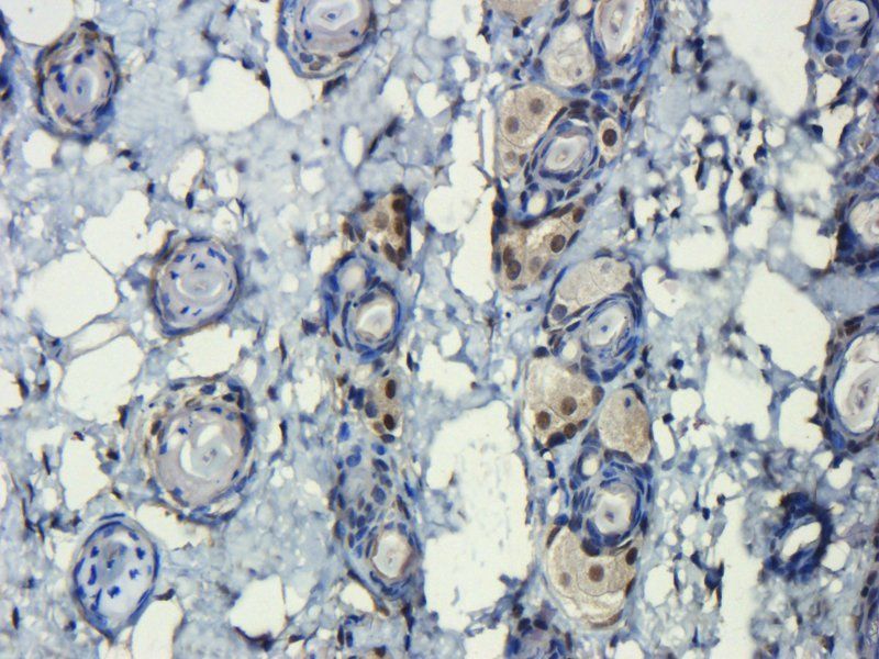
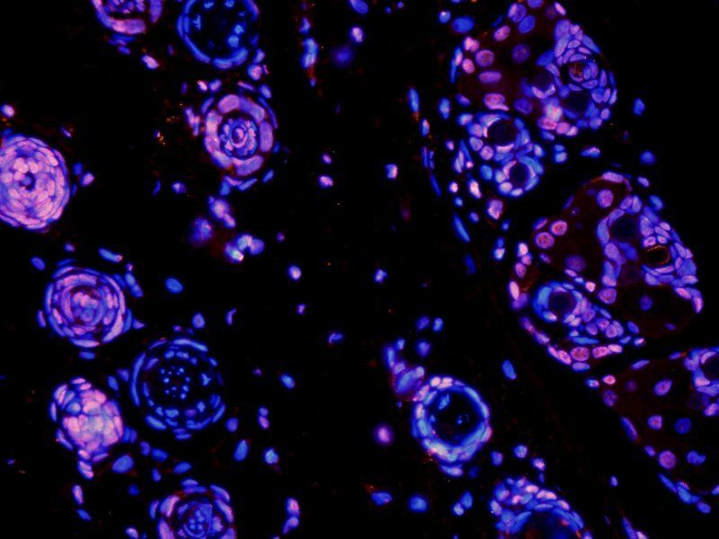
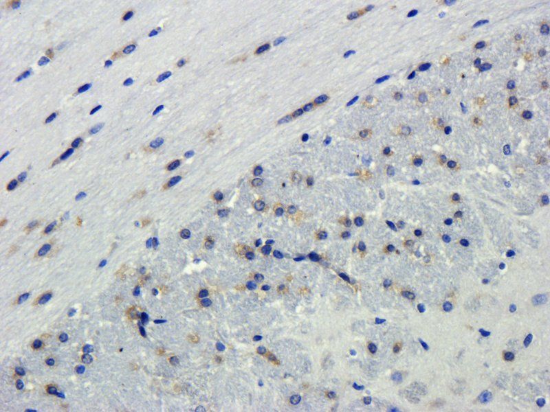
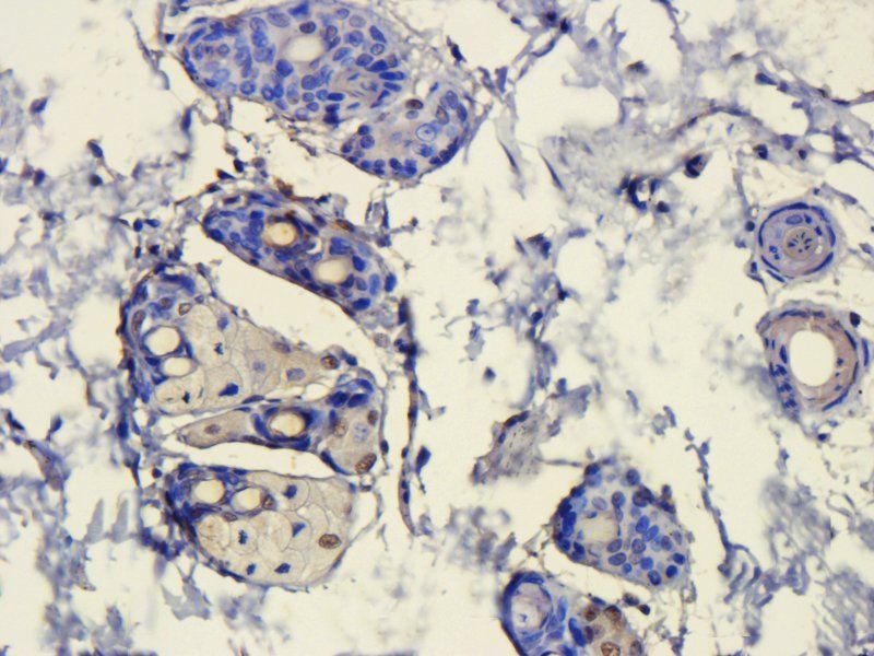
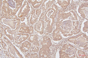
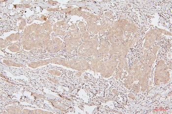
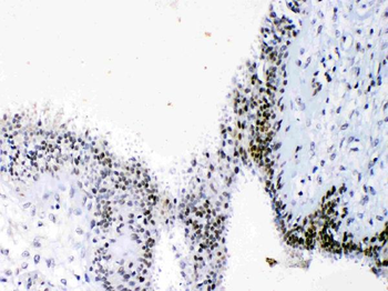
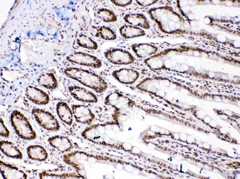
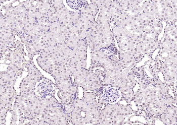
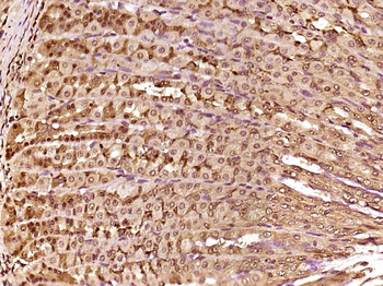
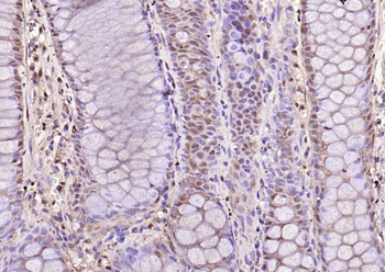
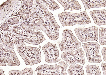
Immunohistochemistry Validation of HMGB1 in Human Stomach Tissue. Immunohistochemical analysis of paraffin-embedded human stomach tissue using anti-HMGB1 antibody (orb1239440) at 1 µg/ml. Tissue was fixed with formaldehyde and blocked with 10% serum for 1 h at RT; antigen retrieval was by heat mediation with a citrate buffer (pH6). Samples were incubated with primary antibody overnight at 4°C. A goat anti-rabbit IgG H&L (HRP) at 1/250 was used as secondary. Counter stained with Hematoxylin.
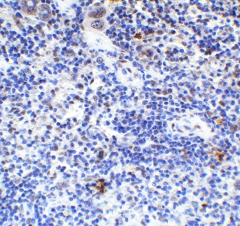
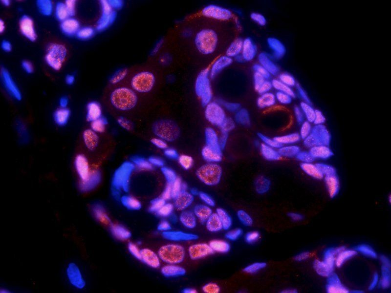
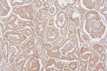
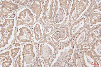
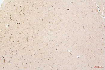
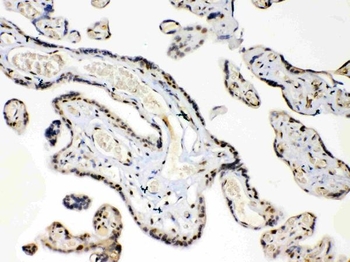
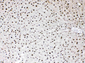
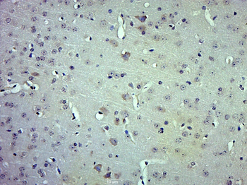
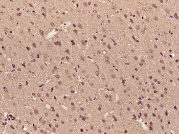
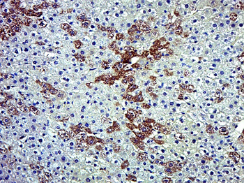
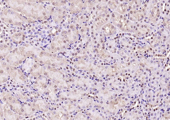
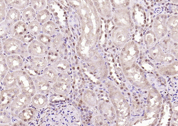
Immunohistochemistry Validation of HMGB1 in Mouse Spleen Tissue. Immunohistochemical analysis of paraffin-embedded mouse spleen tissue using anti-HMGB1 antibody (orb1239440) at 2 µg/ml. Tissue was fixed with formaldehyde and blocked with 10% serum for 1 h at RT; antigen retrieval was by heat mediation with a citrate buffer (pH6). Samples were incubated with primary antibody overnight at 4°C. A goat anti-rabbit IgG H&L (HRP) at 1/250 was used as secondary. Counter stained with Hematoxylin.
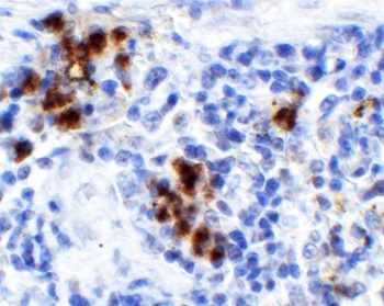
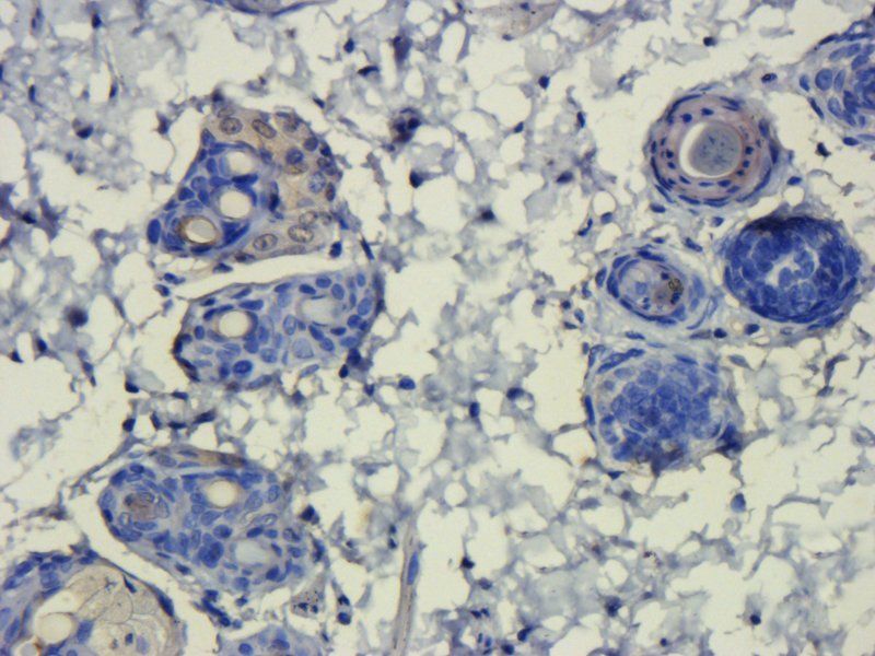
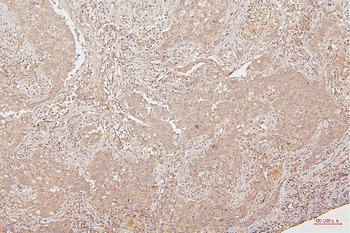
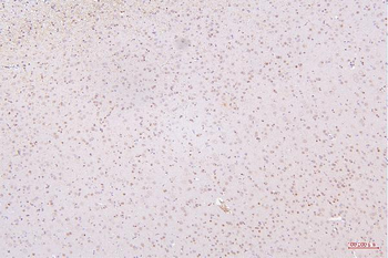
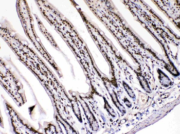
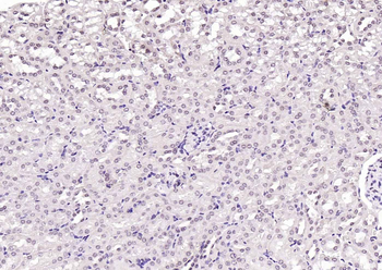
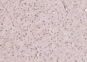
Immunohistochemistry Validation of HMGB1 in Rat Spleen Tissue. Immunohistochemical analysis of paraffin-embedded rat spleen tissue using anti-HMGB1 antibody (orb1239440) at 1 µg/ml. Tissue was fixed with formaldehyde and blocked with 10% serum for 1 h at RT; antigen retrieval was by heat mediation with a citrate buffer (pH6). Samples were incubated with primary antibody overnight at 4°C. A goat anti-rabbit IgG H&L (HRP) at 1/250 was used as secondary. Counter stained with Hematoxylin.
Documents Download
Datasheet
Product Information
Request a Document
Protocol Information
WB
Western Blot (IB, immunoblot)
IHC-P
Immunohistochemistry Paraffin
IF
Immunofluorescence
ELISA
Enzyme-linked Immunosorbent Assay (EIA)
HMGB1 Antibody (orb1239440)

- 0.0
Based on 0 reviews
Participating in our Biorbyt product reviews program enables you to support fellow scientists by sharing your firsthand experience with our products.
Login to Submit a ReviewAvailable Sizes
Select a size below