You have no items in your shopping cart.
Cart summary
Item 1 of 3
Item 1 of 3
HEF1 antibody
Catalog Number: orb344431

| Catalog Number | orb344431 |
|---|---|
| Category | Antibodies |
| Description | HEF1 antibody |
| Species/Host | Mouse |
| Clonality | Monoclonal |
| Clone Number | 2G9 |
| Tested applications | ELISA, IF, IP, WB |
| Reactivity | Human, Mouse, Rat |
| Isotype | IgG1 |
| Immunogen | Anti-HEF1 monoclonal antibody was produced by repeated immunizations with a synthetic peptide corresponding to amino acid residues 82-398 of human HEF1 protein (hHEF1, 843 aa, predicted MW 92.8 kDa). |
| Concentration | 1.0 mg/mL |
| Dilution range | ELISA: 1:5,000 - 1:20,000, IF: 1:500, IP: 1:1,000, WB: 1:5,000 |
| Form/Appearance | Liquid (sterile filtered) |
| Purity | This Protein A purified antibody is directed against human HEF1 protein. The product was purified from tissue culture supernatant by chromatography. Reactivity occurs against human, mouse and rat forms of the protein. Reactivity against multiple isoforms is expected. Reactivity against homologues from other sources is not known. Specificity was determined by partial epitope mapping. |
| Conjugation | Unconjugated |
| UniProt ID | Q14511 |
| NCBI | 5453680 |
| Storage | Store vial at -20° C prior to opening. Aliquot contents and freeze at -20° C or below for extended storage. Avoid cycles of freezing and thawing. Centrifuge product if not completely clear after standing at room temperature. This product is stable for several weeks at 4° C as an undiluted liquid. Dilute only prior to immediate use. |
| Buffer/Preservatives | 0.01% (w/v) Sodium Azide |
| Alternative names | mouse anti-hEF1 antibody, mouse anti-NEDD-9 antibo Read more... |
| Note | For research use only |
| Application notes | This monoclonal antibody has been tested for use in western blotting, immunoprecipitation and immunofluorescence. Specific conditions for reactivity should be optimized by the end user. Expect bands approximately 115 and 105 in size corresponding to isoforms of HEF1 protein by western blotting in the appropriate cell lysate or extract. This antibody does not recognize p130Cas. Sin1 has not been tested. IF was performed using 4% PFA fixed cells. This monoclonal antibody mostly detects HEF1 localized at the focal adhesion sites. |
| Expiration Date | 12 months from date of receipt. |
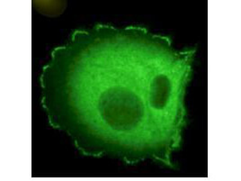
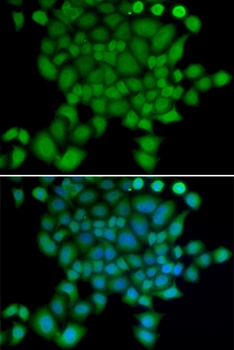
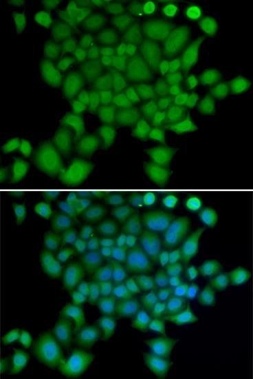
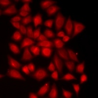
Immunofluorescence microscopy using Biorbyt's Monoclonal anti-HEF1 antibody (clone 2G9) shows detection of HEF1 localized at focal adhesion sites. The antibody was used at a 1:500 dilution with a 3-sec exposure time.
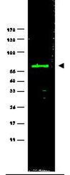
Western blot using Biorbyt's monoclonal anti-HEF1 antibody (clone 2G9) antibody shows detection of a ~92 kDa band corresponding to HEF1 in MCF7 lysate (p/n orb348664) [arrowhead]. Approximately 35 µg of lysate was loaded for SDS-PAGE followed by transfer onto nitrocellulose and reaction with a 1:1000 dilution of anti-HEF1 antibody. Detection occurred using a 1:5000 dilution of IRDye®800 conjugated Sh-a-Mouse IgG [H&L] for 45 min at room temperature (800 nm channel, green). Molecular weight estimation was made by comparison to prestained MW markers (indicated at left).
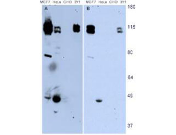
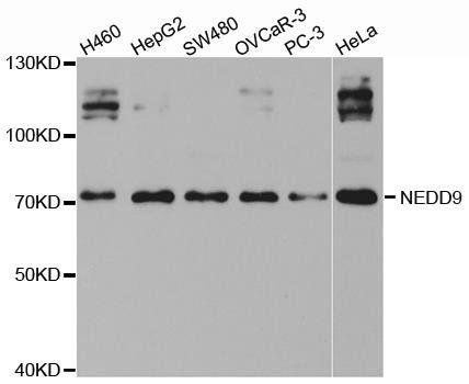
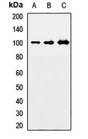
Western blotting using Biorbyt's monoclonal anti-HEF1 antibody (clone 2G9) shows detection of endogenous HEF1 present in various cell lines [MCF7, HeLa, CHO, 3Y1]. Panel A shows detection using a 15 min exposure. Panel B is the same blot exposed for 2 min. The doublet represents p105 and p115 staining. The lower MW band represents p55. 3Y1 cells are derived from rat fibroblast cells. Mouse 3T3 cells are also reactive (not shown). To date no staining has been noted on CHO cells.
- Item 1 of 3
- Item 1 of 3
NEDD-9/CASL/HEF1 Antibody [orb1733239]
IP, WB
Human, Mouse
Rabbit
Recombinant
Unconjugated
20 μl (2 blots) - Item 1 of 3
NEDD-9/CASL/HEF1 Antibody [orb1733240]
IP, WB
Human, Mouse
Rabbit
Recombinant
Unconjugated
100 μl (10 blots) - Item 1 of 3
NEDD9 antibody [orb167180]
ICC, IF, IHC, WB
Human, Mouse, Rat
Polyclonal
Unconjugated
50 μl, 100 μl, 200 μl - Item 1 of 2
NEDD9 antibody [orb412111]
IF, IH, WB
Human, Mouse, Rat
Rabbit
Polyclonal
Unconjugated
50 μl, 100 μl, 200 μl


Submit a review
Filter by Rating

- 5 stars

- 4 stars
- 3 stars
- 2 stars
- 1 stars
