You have no items in your shopping cart.
Cart summary
Item 1 of 5
Item 1 of 5
GRP78 Antibody: PerCP
Catalog Number: orb151313

| Catalog Number | orb151313 |
|---|---|
| Category | Antibodies |
| Description | Rabbit polyclonal to GRP78 (PerCP). GRP78 is a ubiquitously expressed, 78-kDa glucose regulated protein, and is commonly referred to as an immunoglobin chain binding protein (BiP). The BiP proteins are categorized as stress response proteins because they play an important role in the proper folding and assembly of nascent protein and in the scavenging of misfolded proteins in the endoplasmic reticulum lumen. Translation of BiP is directed by an internal ribosomal entry site (IRES) in the 5' non-translated region of the BiP mRNA. BiP IRES activity increases when cells are heat stressed. GRP78 is also critical for maintenance of cell homeostasis and the prevention of apoptosis. Luo et al. have provided findings that suggest GRP78 is essential for embryonic cell growth and pluripotent cell survival. In terms of diseases, GRP78 has been shown to be a reliable biomarker of hypoglycemia, to serve a neuroprotective function in neurons exposed to glutamate and oxidative stress, and its protein levels are reduced in the brains of Alzheimers patients. Also, the induction of the GRP78 protein that results in severe glucose and oxygen deprivation could possible lead to drug resistance to anti-tumor drugs .. |
| Species/Host | Rabbit |
| Clonality | Polyclonal |
| Tested applications | ELISA, ICC, IF, IHC, WB |
| Reactivity | Bovine, Canine, Frog, Fungi, Hamster, Human, Monkey, Mouse, Plant, Rabbit, Rat |
| Immunogen | Rat GRP78 (Bip) synthetic peptide conjugated to KLH |
| Concentration | 1 mg/ml |
| Dilution range | WB (1:1000), ICC/IF (1:100) |
| Conjugation | PerCP |
| MW | 78kDa |
| Target | GRP78 |
| Entrez | 25617 |
| UniProt ID | P06761 |
| NCBI | NP_037215.1 |
| Storage | Conjugated antibodies should be stored according to the product label |
| Buffer/Preservatives | 95.64mM Phosphate, 2.48mM MES and 2mM EDTA |
| Alternative names | 78 kDa glucose regulated protein antibody, 78 kDa Read more... |
| Note | For research use only |
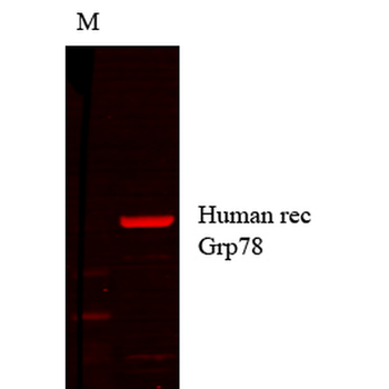
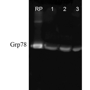
| Application notes | A 1:1000 dilution of SPC-107 was sufficient for detection of Grp78 in 10 µg of rat tissue lysate by ECL immunoblot analysis. |
| Expiration Date | 12 months from date of receipt. |

Immunocytochemistry/Immunofluorescence analysis using Rabbit Anti-GRP78 Polyclonal Antibody. Tissue: Hippocampal Section. Species: Mouse. Fixation: 4% Formaldehyde for 12 hours at RT. Paraffin embedded. Primary Antibody: Rabbit Anti-GRP78 Polyclonal Antibody at 1:100 for 12 hours at 4°C. Secondary Antibody: Alexa Fluor 555 Goat Anti-Rabbit at 1:250 for 1 hour at RT. Counterstain: Hoechst at 1:1000 for 10 min at RT. Localization: Grp78 staining in mouse pyramidal cell layer. Magnification: 20x. Slice thickness: 7 microns.
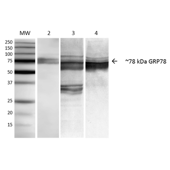
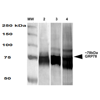
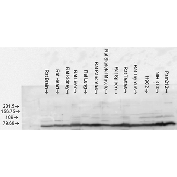
Western blot analysis of Human, Dog, Mouse Cell line lysates showing detection of GRP78 protein using Rabbit Anti-GRP78 Polyclonal Antibody. Primary Antibody: Rabbit Anti-GRP78 Polyclonal Antibody at 1:1000.
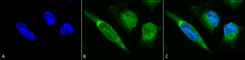
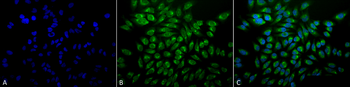
Immunocytochemistry/Immunofluorescence analysis using Rabbit Anti-GRP78 Polyclonal Antibody. Tissue: Heat Shocked Cervical cancer cell line (HeLa). Species: Human. Fixation: 2% Formaldehyde for 20 min at RT. Primary Antibody: Rabbit Anti-GRP78 Polyclonal Antibody at 1:100 for 12 hours at 4°C. Secondary Antibody: FITC Goat Anti-Rabbit (green) at 1:200 for 2 hours at RT. Counterstain: DAPI (blue) nuclear stain at 1:40000 for 2 hours at RT. Localization: Endoplasmic reticulum lumen. Melanosome. Cytoplasm. Magnification: 100x. (A) DAPI (blue) nuclear stain. (B) Anti-GRP78 Antibody. (C) Composite. Heat Shocked at 42°C for 30 min.
Western blot analysis of Rat Tissue lysates showing detection of GRP78 protein using Rabbit Anti-GRP78 Polyclonal Antibody. Load: 15 μgprotein. Block: 1.5% BSA. Primary Antibody: Rabbit Anti-GRP78 Polyclonal Antibody at 1:1000 for 2 hours at RT. Secondary Antibody: Donkey Anti-Rabbit IgG: HRP for 1 hour at RT.
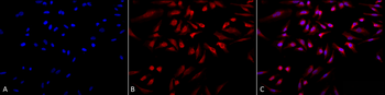
Immunocytochemistry/Immunofluorescence analysis using Rabbit Anti-GRP78 Polyclonal Antibody. Tissue: Heat Shocked Cervical cancer cell line (HeLa). Species: Human. Fixation: 2% Formaldehyde for 20 min at RT. Primary Antibody: Rabbit Anti-GRP78 Polyclonal Antibody at 1:100 for 12 hours at 4°C. Secondary Antibody: APC Goat Anti-Rabbit (red) at 1:200 for 2 hours at RT. Counterstain: DAPI (blue) nuclear stain at 1:40000 for 2 hours at RT. Localization: Endoplasmic reticulum lumen. Melanosome. Cytoplasm. Magnification: 20x. (A) DAPI (blue) nuclear stain. (B) Anti-GRP78 Antibody. (C) Composite. Heat Shocked at 42°C for 30 min.
- Item 1 of 4
GRP78 Antibody: PerCP [orb148012]
ELISA, ICC, IF, IHC, WB
Bovine, Frog, Fungi, Hamster, Human, Monkey, Mouse, Plant, Rabbit, Rat
Mouse
Monoclonal
PerCP
100 μg - Item 1 of 3
GRP78 Antibody: PerCP [orb181923]
ELISA, ICC, IF, IHC, WB
Human, Mouse, Rat
Mouse
Monoclonal
PerCP
100 μg - Item 1 of 3
GRP78 Antibody: PerCP [orb181941]
ELISA, ICC, IF, IHC, WB
Human, Mouse, Rat
Mouse
Monoclonal
PerCP
100 μg - Item 1 of 3
KDEL Antibody: PerCP [orb151347]
ELISA, ICC, IF, IHC, WB
Human, Mouse, Rat
Rabbit
Polyclonal
PerCP
200 μg - Item 1 of 2
GRP78 (Bip) Antibody: PerCP [orb151968]
ELISA, ICC, IF, IHC, WB
Canine, Drosophila, Hamster, Human, Mouse, Rat
Rabbit
Polyclonal
PerCP
100 μg