You have no items in your shopping cart.
Cart summary
Item 1 of 4
Item 1 of 4
GRP78 Antibody
Catalog Number: orb67383

| Catalog Number | orb67383 |
|---|---|
| Category | Antibodies |
| Description | Mouse Anti-Human GRP78 Monoclonal IgG2b |
| Species/Host | Mouse |
| Clonality | Monoclonal |
| Clone Number | 1H11-1H7 |
| Tested applications | ICC, IF, WB |
| Reactivity | Bovine, Frog, Fungi, Hamster, Human, Monkey, Mouse, Plant, Rabbit, Rat |
| Isotype | IgG2b |
| Immunogen | His-tagged human GRP78 |
| Concentration | 1 mg/ml |
| Dilution range | WB (1:2000), ICC/IF (1:100) |
| Conjugation | Unconjugated |
| MW | 78kDa |
| Target | GRP78 |
| Entrez | 14828 |
| UniProt ID | P20029 |
| NCBI | NP_001156906.1 |
| Storage | Maintain refrigerated at 2-8°C for up to 2 weeks. For long term storage store at -20°C in small aliquots to prevent freeze-thaw cycles. |
| Buffer/Preservatives | PBS pH 7.4, 50% glycerol, 0.09% sodium azide *Storage buffer changes when conjugated |
| Alternative names | 78 kDa glucose regulated protein Antibody, 78 kDa Read more... |
| Note | For research use only |
| Application notes | 0.5 µg/ml of SMC-195 was sufficient for detection of Grp78 in 10 µg of rat tissue lysate by ECL immunoblot analysis. |
| Expiration Date | 12 months from date of receipt. |

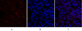
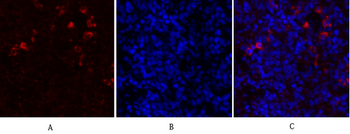
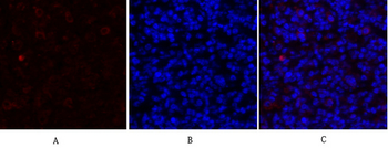
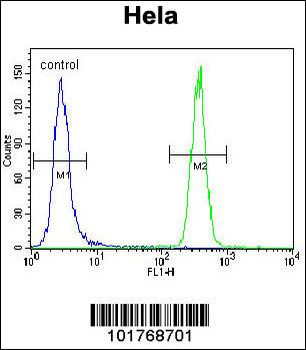
Immunocytochemistry/Immunofluorescence analysis using Mouse Anti-GRP78 Monoclonal Antibody, Clone 1H11-1H7. Tissue: Heat Shocked cervical cancer cells (HeLa). Species: Human. Fixation: 2% Formaldehyde for 20 min at RT. Primary Antibody: Mouse Anti-GRP78 Monoclonal Antibody at 1:100 for 12 hours at 4°C. Secondary Antibody: FITC Goat Anti-Mouse (green) at 1:200 for 2 hours at RT. Counterstain: DAPI (blue) nuclear stain at 1:40000 for 2 hours at RT. Localization: Endoplasmic reticulum lumen. Melanosome. Magnification: 100x. (A) DAPI (blue) nuclear stain. (B) Anti-GRP78 Antibody. (C) Composite. Heat Shocked at 42°C for 1h.
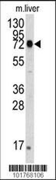
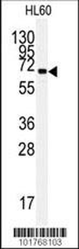
Western Blot analysis of Human cell lysates showing detection of GRP78 protein using Mouse Anti-GRP78 Monoclonal Antibody, Clone 1H11-1H7. Primary Antibody: Mouse Anti-GRP78 Monoclonal Antibody at 1:1000.
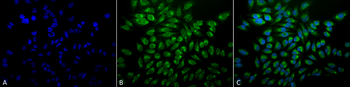
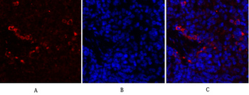
Immunocytochemistry/Immunofluorescence analysis using Mouse Anti-GRP78 Monoclonal Antibody, Clone 1H11-1H7. Tissue: Heat Shocked cervical cancer cells (HeLa). Species: Human. Fixation: 2% Formaldehyde for 20 min at RT. Primary Antibody: Mouse Anti-GRP78 Monoclonal Antibody at 1:100 for 12 hours at 4°C. Secondary Antibody: FITC Goat Anti-Mouse (green) at 1:200 for 2 hours at RT. Counterstain: DAPI (blue) nuclear stain at 1:40000 for 2 hours at RT. Localization: Endoplasmic reticulum lumen. Melanosome. Magnification: 20x. (A) DAPI (blue) nuclear stain. (B) Anti-GRP78 Antibody. (C) Composite. Heat Shocked at 42°C for 1h.
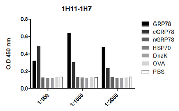
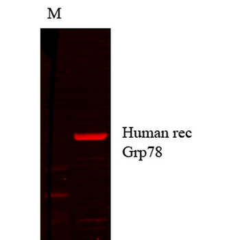
ELISA analysis using Mouse Anti-GRP78 Monoclonal Antibody, Clone 1H11-1H7. Primary Antibody: Mouse Anti-GRP78 Monoclonal Antibody. Secondary Antibody: Goat anti-mouse IgG: HRP at 1:10000.
- Item 1 of 8
- Item 1 of 7
HSP A5 Polyclonal Antibody [orb1413406]
IF, IHC-P, WB
Human, Mouse, Rat
Rabbit
Polyclonal
Unconjugated
100 μl - Item 1 of 5
- Item 1 of 6
- Item 1 of 5
HSPA5 Antibody [orb676526]
ELISA, IHC, WB
Human, Mouse, Rat
Rabbit
Polyclonal
Unconjugated
50 μg, 100 μg