You have no items in your shopping cart.
Cart summary
Item 1 of 7
Item 1 of 7
GPX1 Antibody
Catalog Number: orb1262271

| Catalog Number | orb1262271 |
|---|---|
| Category | Antibodies |
| Description | GPX1 Antibody |
| Species/Host | Rabbit |
| Clonality | Polyclonal |
| Tested applications | FC, IF, IHC-P, WB |
| Reactivity | Human, Mouse, Rat |
| Isotype | Rabbit Ig |
| Immunogen | This GPX1 antibody is generated from rabbits immunized with a KLH conjugated synthetic peptide between 164-193 amino acids from the C-terminal region of human GPX1. |
| Antibody Type | Primary Antibody |
| Concentration | batch dependent |
| Form/Appearance | Liquid |
| Conjugation | Unconjugated |
| MW | 22 kDa |
| Target | GPX1 |
| UniProt ID | P07203 |
| NCBI | P07203 |
| Storage | Maintain refrigerated at 2-8°C for up to 2 weeks. For long term storage store at -20°C in small aliquots to prevent freeze-thaw cycles. |
| Buffer/Preservatives | Supplied in PBS with 0.09% (W/V) sodium azide. |
| Alternative names | Glutathione peroxidase 1, GPx-1, GSHPx-1, Cellular Read more... |
| Note | For research use only |
| Application notes | For FACS starting dilution is: 1:25For WB starting dilution is: 1:2000 |
| Expiration Date | 12 months from date of receipt. |

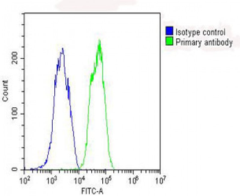
Overlay histogram showing HepG2 cells stained with Antibody (green line). The cells were fixed with 2% paraformaldehyde (10 min) and then permeabilized with 90% methanol for 10 min. The cells were then icubated in 2% bovine serum albumin to block non-specific protein-protein interactions followed by the antibody (1:25 dilution) for 60 min at 37°C. The secondary antibody used was Goat-Anti-Rabbit IgG, Conjugated Highly Cross-Adsorbed at 1/200 dilution for 40 min at 37°C. Isotype control antibody (blue line) was rabbit IgG (1ug/1x10^6 cells) used under the same conditions. Acquisition of > 10000 events was performed.

Overlay histogram showing HepG2 cells stained with Antibody (green line). The cells were fixed with 2% paraformaldehyde (10 min) and then permeabilized with 90% methanol for 10 min. The cells were then icubated in 2% bovine serum albumin to block non-specific protein-protein interactions followed by the antibody (1:25 dilution) for 60 min at 37°C. The secondary antibody used was Goat-Anti-Rabbit IgG, Conjugated Highly Cross-Adsorbed at 1/200 dilution for 40 min at 37°C. Isotype control antibody (blue line) was rabbit IgG (1ug/1x10^6 cells) used under the same conditions. Acquisition of > 10000 events was performed.

Overlay histogram showing HepG2 cells stained with Antibody (green line). The cells were fixed with 2% paraformaldehyde (10 min) and then permeabilized with 90% methanol for 10 min. The cells were then icubated in 2% bovine serum albumin to block non-specific protein-protein interactions followed by the antibody (1:25 dilution) for 60 min at 37°C. The secondary antibody used was Goat-Anti-Rabbit IgG, Conjugated Highly Cross-Adsorbed at 1/200 dilution for 40 min at 37°C. Isotype control antibody (blue line) was rabbit IgG (1ug/1x10^6 cells) used under the same conditions. Acquisition of > 10000 events was performed.

Overlay histogram showing HepG2 cells stained with Antibody (green line). The cells were fixed with 2% paraformaldehyde (10 min) and then permeabilized with 90% methanol for 10 min. The cells were then icubated in 2% bovine serum albumin to block non-specific protein-protein interactions followed by the antibody (1:25 dilution) for 60 min at 37°C. The secondary antibody used was Goat-Anti-Rabbit IgG, Conjugated Highly Cross-Adsorbed at 1/200 dilution for 40 min at 37°C. Isotype control antibody (blue line) was rabbit IgG (1ug/1x10^6 cells) used under the same conditions. Acquisition of > 10000 events was performed.

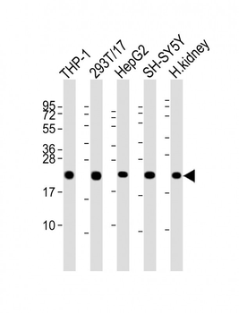
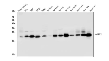
Western Blot at 1:2000 dilution Lane 1: THP-1 whole cell lysate Lane 2: 293T/17 whole cell lysate Lane 3: HepG2 whole cell lysate Lane 4: SH-SY5Y whole cell lysate Lane 5: human kidney lysate Lysates/proteins at 20 ug per lane.

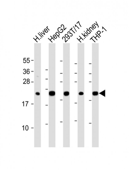
Western Blot at 1:2000 dilution Lane 1: human liver lysate Lane 2: HepG2 whole cell lysate Lane 3: 293T/17 whole cell lysate Lane 4: human kidney lysate Lane 5: THP-1 whole cell lysate Lysates/proteins at 20 ug per lane.

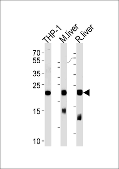
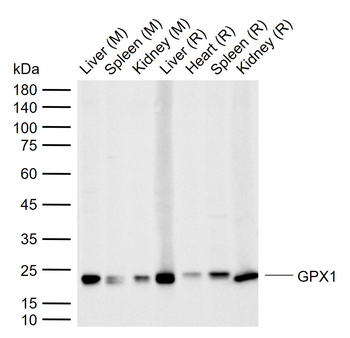
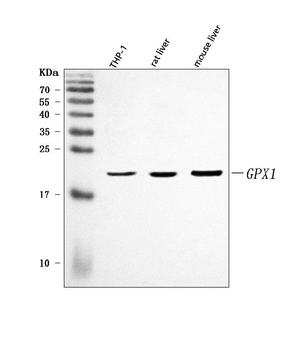
Western blot analysis of lysates from THP-1 cell line, mouse liver and rat liver tissue (from left to right), using GPX1 Antibody.
- Item 1 of 7
GPX1 Antibody (C-term) [orb1927986]
FC, IF, IHC-P, WB
Human, Mouse, Rat
Rabbit
Polyclonal
Unconjugated
400 μl, 80 μl - Item 1 of 4
Goat anti-Glutathione Peroxidase 1 (isoform1) Antibody [orb19249]
ELISA, IHC, WB
Human
Goat
Polyclonal
Unconjugated
100 μg - Item 1 of 6
Anti-GPX1 Antibody (monoclonal, 8B10) [orb527061]
FC, ICC, IF, IHC, WB
Human, Mouse, Rat
Mouse
Monoclonal
Unconjugated
10 μg - Item 1 of 3
GPX1 Rabbit Polyclonal Antibody [orb5344]
ELISA, FC, IF, IHC-Fr, IHC-P, WB
Bovine, Canine, Equine, Guinea pig, Porcine
Human, Mouse, Rat, Sheep
Rabbit
Polyclonal
Unconjugated
100 μl, 50 μl - Item 1 of 4
Anti-GPX1 Antibody [orb1728187]
ELISA, IHC, WB
Human, Mouse, Rat
Rabbit
Polyclonal
Unconjugated
10 μg, 100 μg