You have no items in your shopping cart.
Cart summary
Item 1 of 3
Item 1 of 3
GPD1L Antibody
Catalog Number: orb1270636

| Catalog Number | orb1270636 |
|---|---|
| Category | Antibodies |
| Description | GPD1L Antibody |
| Species/Host | Rabbit |
| Clonality | Polyclonal |
| Tested applications | FC, IHC-P, WB |
| Reactivity | Human, Mouse |
| Isotype | Rabbit Ig |
| Immunogen | This GPD1L antibody is generated from rabbits immunized with a KLH conjugated synthetic peptide between 44-73 amino acids from the N-terminal region of human GPD1L. |
| Concentration | batch dependent |
| Dilution range | For WB starting dilution is: 1:1000For IHC-P starting dilution is: 1:50~100For FACS starting dilution is: 1:10~50 |
| Form/Appearance | Liquid |
| Conjugation | Unconjugated |
| MW | 38 kDa |
| Target | GPD1L |
| UniProt ID | Q8N335 |
| NCBI | Q8N335 |
| Storage | Store at 4°C for three months and -20°C, stable for up to one year. As with all antibodies care should be taken to avoid repeated freeze thaw cycles. Antibodies should not be exposed to prolonged high temperatures. |
| Buffer/Preservatives | Supplied in PBS with 0.09% (W/V) sodium azide. |
| Alternative names | Glycerol-3-phosphate dehydrogenase 1-like protein, Read more... |
| Note | For research use only |
| Application notes | For WB starting dilution is: 1:1000For IHC-P starting dilution is: 1:50~100For FACS starting dilution is: 1:10~50 |
| Expiration Date | 12 months from date of receipt. |

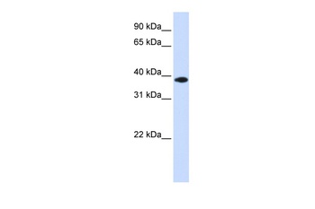
Western Blot at 1:1000 dilution Lane 1: MCF-7 whole cell lysate Lane 2: human heart lysate Lysates/proteins at 20 ug per lane.

GPD1L antibody immunohistochemistry analysis in formalin fixed and paraffin embedded mouse heart tissue followed by peroxidase conjugation of the secondary antibody and DAB staining.

Flow cytometric analysis of MCF-7 cells (right histogram) compared to a negative control cell (left histogram). FITC-conjugated goat-anti-rabbit secondary antibodies were used for the analysis.
- Item 1 of 4
- Item 1 of 2
GPD1L antibody [orb183880]
ELISA, ICC, IF, IHC-Fr, IHC-P
Bovine, Canine, Gallus, Human, Mouse, Sheep
Rat
Rabbit
Polyclonal
Unconjugated
50 μl, 100 μl, 200 μl - Item 1 of 1
- Item 1 of 1
- Item 1 of 1


Submit a review
Filter by Rating

- 5 stars

- 4 stars
- 3 stars
- 2 stars
- 1 stars
