You have no items in your shopping cart.
Goat IgG (H&L) Antibody Fluorescein Conjugated Pre-Adsorbed
Catalog Number: orb347030
| Catalog Number | orb347030 |
|---|---|
| Category | Antibodies |
| Description | Goat IgG (H&L) antibody (FITC) |
| Species/Host | Donkey |
| Clonality | Polyclonal |
| Tested applications | FC, FLISA, IF |
| Reactivity | Goat |
| Isotype | IgG |
| Immunogen | Goat IgG whole molecule |
| Concentration | 1.0 mg/mL |
| Dilution range | FLISA: 1:10,000 - 1:50,000, FC: 1:500 - 1:2,500, IF: 1:1,000 - 1:5,000 |
| Form/Appearance | Lyophilized |
| Purity | This product was prepared from monospecific antiserum by immunoaffinity chromatography using Goat IgG coupled to agarose beads followed by solid phase adsorption(s) to remove any unwanted reactivities. Assay by immunoelectrophoresis resulted in a single precipitin arc against anti-Fluorescein, anti-Donkey Serum, Goat IgG and Goat Serum. No reaction was observed against Chicken, Guinea Pig, Hamster, Horse, Mouse, Rabbit and Rat Serum Proteins. |
| Conjugation | FITC |
| Storage | Store vial at 4° C prior to restoration. For extended storage aliquot contents and freeze at -20° C or below. Avoid cycles of freezing and thawing. Centrifuge product if not completely clear after standing at room temperature. This product is stable for several weeks at 4° C as an undiluted liquid. Dilute only prior to immediate use. |
| Buffer/Preservatives | 0.01% (w/v) Sodium Azide |
| Alternative names | Donkey anti-Goat IgG Antibody Fluorescein conjugat Read more... |
| Note | For research use only |
| Application notes | This product is designed for immunofluorescence microscopy, fluorescence based plate assays (FLISA) and fluorescent western blotting. This product is also suitable for multiplex analysis, including multicolor imaging, utilizing various commercial platforms. |
| Expiration Date | 12 months from date of receipt. |

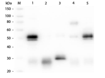
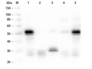
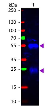
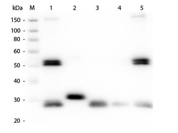
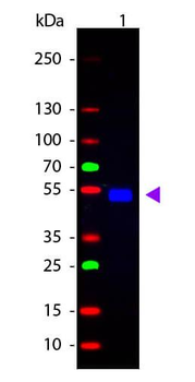
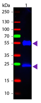
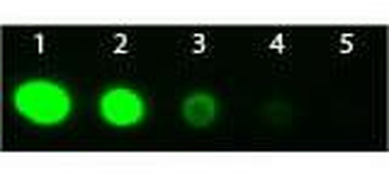
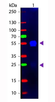
Western Blot results using Donkey Anti-Goat IgG FITC. Mature cathepsins K, L, S, and V are zymographically active and migrate at distinct electrophoretic distances A) Recombinant cathepsins K, S, and V (1, 20, and 50 ng) from E. coli and cathepsin L (50 ng) isolated from human liver were loaded for cathepsin gelatin zymography and incubated overnight in acetate buffer, pH6. The zymogram reveals zymographically active bands at different electrophoretic migration distances. B) Mature, recombinant cathepsins K, S, and V (10 ng) from eukaryotic expression systems and cathepsin L (50 ng) isolated from human liver were loaded separately and all in one lane (where indicated) for gelatin zymography assayed at pH6. C) Western blot analysis of 50 ng of recombinant glycosylated cathepsin K, L, S, and V from eukaryotic expression systems also were loaded for non-reduced Western blotting. D) Monocyte-derived macrophages and monocyte-derived osteoclasts were lysed and equal amounts of protein were loaded for cathepsin zymography and E) reduced, fully denaturing Western blotting for cathepsins K and V. Procathepsin (pro) bands are at ~37 kDa and mature (mat) cathepsin bands are at ~27 kDa. Increased cathepsins K and V were detected in the osteoclasts compared to the macrophages.
Rabbit IgG (H&L) Secondary Antibody Fluorescein Conjugated Pre-Adsorbed [orb347650]
DOT, FC, FLISA, IF, WB
Rabbit
Goat
Polyclonal
FITC
1 mgRabbit IgG (H&L) Antibody Fluorescein Conjugated Pre-Adsorbed [orb347670]
IF, WB
Rabbit
Goat
Polyclonal
FITC
2 mgRat IgG (H&L) Antibody Fluorescein Conjugated Pre-Adsorbed [orb347787]
FC, FLISA, IF, WB
Rat
Goat
Polyclonal
FITC
1 mgMouse IgG (H&L) Secondary Antibody Fluorescein Conjugated Pre-Adsorbed [orb347377]
DOT, FC, FLISA, IF, WB
Mouse
Goat
Polyclonal
FITC
1 mgF(ab')2 Rabbit IgG (H&L) Antibody Fluorescein Conjugated Pre-Adsorbed [orb348305]
DOT, FC, FLISA, IF, WB
Rabbit
Goat
Polyclonal
FITC
500 μg


